




DcR3 recombinant protein: a key molecule in sepsis inflammation regulation and multi-organ protection
Author: Senior Product Manager of UA Views:7 


This article focuses on the molecular characteristics and biological functions of decoy receptor 3 (DcR3), systematically elaborating its protective role as a potential biomarker for inflammatory diseases in sepsis, analyzing the molecular mechanisms by which DcR3 improves sepsis prognosis through inhibiting inflammatory responses and cell apoptosis, and regulating immune cell composition and gut microbiota, and exploring the therapeutic value of DcR3 in multi-organ protection of the lungs, liver, and kidneys as well as in the maintenance of the intestinal barrier.
- Recent Advances
- Product Information
Recent Advances
DcR3 Recombinant Protein: A Key Molecule in Sepsis Inflammation Regulation and Multi-Organ Protection
Summary: This article focuses on the molecular characteristics and biological functions of decoy receptor 3 (DcR3), systematically elaborating its protective role in sepsis as a potential biomarker for inflammatory diseases, analyzing the molecular mechanisms by which DcR3 improves sepsis prognosis through inhibiting inflammatory responses and cell apoptosis, regulating immune cell composition and gut microbiota, and discussing the therapeutic value of DcR3 in multi-organ protection of the lungs, liver, and kidneys as well as intestinal barrier maintenance.
I. The Therapeutic Dilemma of Sepsis and the Research Background of DcR3.
Despite the high mortality rate of sepsis, there is a lack of specific drugs for treating sepsis. Decoy receptor 3 (DcR3) is a potential biomarker for the progression of inflammatory diseases. Recombinant human DcR3-Fc chimeric protein can inhibit the inflammatory response in septic mice, which is crucial for improving survival rates. However, the Fc region may have adverse effects on patients, while endogenous polypeptides are highly favorable for clinical application. However, the mechanism by which DcR3 affects sepsis remains unclear. Against this background, researchers aimed to demonstrate that DcR3 may be beneficial for treating sepsis and to investigate its mechanism of action. The researchers obtained recombinant DcR3 in vitro and performed post-operative DcR3 treatment in lipopolysaccharide- and cecal ligation and puncture-induced septic mouse models to explore its potential molecular mechanisms. DcR3 inhibited persistent excessive inflammatory responses in vitro, improved survival rates, reduced pro-inflammatory cytokine levels, altered circulating immune cell composition, regulated gut microbiota, and induced short-chain fatty acid synthesis in vivo. Therefore, DcR3 can prevent cecal ligation and puncture-induced sepsis by inhibiting inflammatory responses and cell apoptosis. This study provides valuable insights into the molecular mechanisms related to the protective effects of DcR3 against sepsis, paving the way for future clinical research.
II. Regulation of Macrophage Inflammatory Cytokine Expression by DcR3.
The purified DcR3 was verified by SDS-PAGE with a molecular weight of approximately 33 kDa. The results showed that DcR3 had negligible cytotoxicity on RAW264.7 cells within the concentration range of 0 to 5.0 μg/μL. Furthermore, DcR3 at a concentration of 1.0 μg/μL significantly inhibited lipopolysaccharide-induced upregulation of IL-1β, IL-6, and TNF-α in RAW264.7 cells. This in vitro experimental result established that DcR3 has a direct inhibitory effect on excessive inflammatory responses in macrophages.
III. Therapeutic Effects of DcR3 on Lipopolysaccharide- and CLP-Induced Septic Mice.
The researchers first injected DcR3 into sham-operated mice to evaluate its effects on survival rates and inflammatory marker expression, and the results showed that DcR3 had no significant effects on these indicators in sham-operated mice. The therapeutic effects of DcR3 and DcR3.Fc were evaluated using a lipopolysaccharide-induced septic mouse model, and both significantly reduced pro-inflammatory factors.
Subsequently, the researchers used a cecal ligation and puncture-induced septic mouse model to determine the therapeutic potential of DcR3. Compared with the untreated cecal ligation and puncture group, the DcR3 treatment group showed reduced agglutination, improved physical condition, activity, and appetite. DcR3 treatment significantly reduced the sepsis score in cecal ligation and puncture-induced septic mice, the survival rate of the DcR3 treatment group was higher than that of the control group, and the body temperature of mice in the DcR3 treatment group rapidly returned to normal. These findings suggest that DcR3 may play an important therapeutic role in cecal ligation and puncture-induced septic mice.
IV. Inhibitory Effects of DcR3 on Multi-Organ Inflammatory Cytokines and Pathological Damage.
To investigate the role of DcR3 in regulating inflammatory cytokine expression in vivo, the researchers examined whether DcR3 inhibited cecal ligation and puncture-induced inflammatory cytokine levels in serum, heart, liver, spleen, lung, and kidney tissues. Enzyme-linked immunosorbent assay results showed that DcR3 treatment significantly reduced the cecal ligation and puncture-induced elevation of IL-1β, IL-6, and TNF-α levels in serum at 12 and 24 hours, with similar results obtained in heart, liver, spleen, lung, and kidney tissues. Furthermore, reverse transcription quantitative PCR was used to study the inhibitory effects of DcR3 on inflammatory cytokine mRNA expression in heart, liver, spleen, lung, and kidney tissues, and the inflammatory cytokine expression levels in the DcR3 treatment group were significantly lower than those in the cecal ligation and puncture group.
At the pathological level, DcR3 treatment alleviated lung injury, alveolar wall thickening, and hemorrhage in mice; in the liver, the DcR3 treatment group showed less liver damage, reduced inflammatory cells, and reduced swelling; after DcR3 treatment, kidney damage was alleviated, the degree of glomerular rupture was reduced, and glomerular shape was regular. These findings indicate that DcR3 can significantly ameliorate cecal ligation and puncture-induced damage to lung, liver, and kidney tissues in mice.
V. Regulatory Effects of DcR3 on Circulating Immune Cells and the Intestinal Barrier.
Through quantitative detection of peripheral blood from cecal ligation and puncture-induced septic mice, the researchers found that the white blood cell count in the DcR3 treatment group was significantly lower than that in the cecal ligation and puncture group, similar to the sham-operated group. Compared with the cecal ligation and puncture group, monocytes, granulocytes, and lymphocytes were significantly reduced in the DcR3 treatment group. Twelve hours after DcR3 administration, the number of activated NK cells in the DcR3 combined with cecal ligation and puncture group was significantly lower than that in the cecal ligation and puncture group. These findings indicate that DcR3 altered activated NK cells in cecal ligation and puncture-induced septic mice.
In terms of intestinal barrier protection, the colon tissue of sham-operated mice remained normal, with orderly crypts and intact epithelial cells, while the colon tissue of cecal ligation and puncture group mice showed severe damage and increased permeability, which was significantly alleviated after DcR3 treatment. The study emphasized the role of tight junction proteins Occludin and Claudin-1, and DcR3 reversed the effects of cecal ligation and puncture by significantly upregulating the mRNA and protein levels of these proteins, demonstrating a dual protective effect. Therefore, this study revealed the dual mechanism of action of DcR3, which can effectively combat sepsis-induced intestinal barrier dysfunction at both mRNA and protein expression levels.
VI. Regulation of Gut Microbiota Balance and Short-Chain Fatty Acid Production by DcR3.
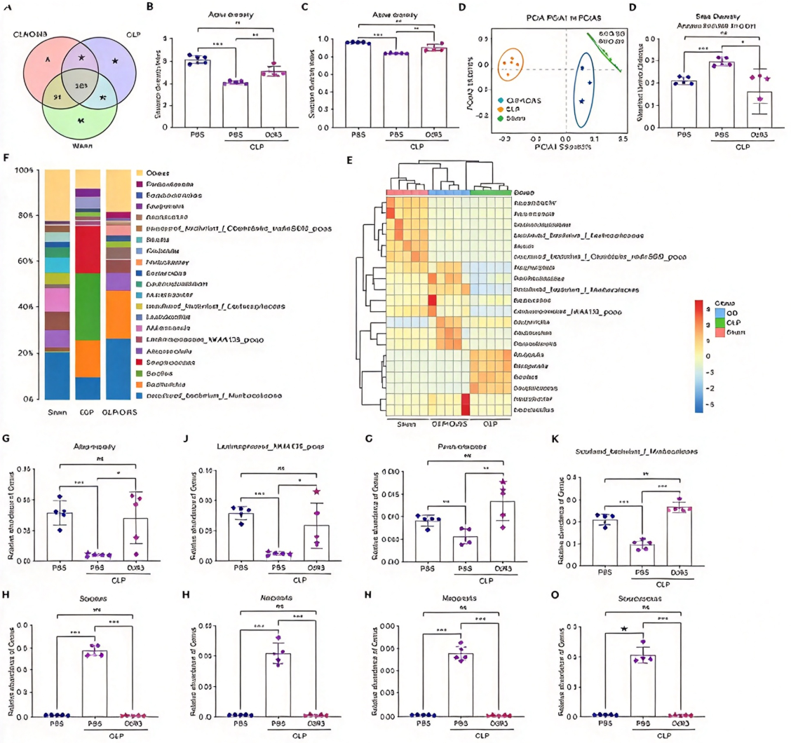
Compared with the cecal ligation and puncture group, the DcR3 treatment group showed significant differences in α diversity indices, and principal coordinate analysis revealed significant differences in gut microbial communities among the three groups. At the genus level, compared with the cecal ligation and puncture group, the abundance of beneficial bacteria was significantly increased in the DcR3 treatment group, while the numbers of pathogenic bacteria such as Bacillus, Klebsiella, Morganella, and Streptococcus were significantly reduced. Therefore, DcR3 can effectively reduce the number of pathogenic bacteria, increase the number of beneficial bacteria, and significantly improve gut dysbiosis in septic mice.
As major metabolites of gut microbiota, short-chain fatty acids play crucial roles in inflammation and immunity. After DcR3 treatment, acetic acid and propionic acid were significantly reduced compared with the cecal ligation and puncture group, while butyric acid was significantly increased after treatment. Correlation analysis showed that Escherichia and Streptococcus were significantly positively correlated with acetic acid and propionic acid, while Blautia, Lachnospiraceae NK4A136 group, and Akkermansia were significantly negatively correlated with these two short-chain fatty acids. Furthermore, the expression of IL-1β, IL-6, and TNF-α in serum and lung tissues was positively correlated with the numbers of Klebsiella, Morganella, and Bacillus. These results indicate that DcR3 exerts anti-inflammatory effects by regulating gut microbiota and short-chain fatty acid metabolism.
VII. Molecular Mechanisms of DcR3 in Inhibiting Inflammatory Responses and Cell Apoptosis.
To elucidate the mechanistic role of DcR3 in sepsis defense, the researchers performed whole-genome RNA sequencing. Notably, DcR3 reduced the activity of key genes in multiple pathways, including NF-κB, TNF, MAPK/ERK, phosphatidylinositol system, and apoptosis-related pathways. Gene Ontology and Kyoto Encyclopedia of Genes and Genomes analyses showed that cecal ligation and puncture significantly enhanced cytokine interactions and TNF, phosphatidylinositol, NOD-like receptor, and IL-17 signaling pathways, while DcR3 effectively inhibited inflammatory responses, disrupted tight junctions, and alleviated apoptotic processes. DcR3 showed strong affinity for competitive binding with FasL, LIGHT, and TL1A, effectively inhibiting downstream signaling pathways triggered by cecal ligation and puncture. Western blot showed that DcR3 significantly attenuated NF-κB activation marked by increased phosphorylation of NF-κB p65 and IKBα in cecal ligation and puncture, and the ratio of cleaved caspase-3 was significantly reduced after DcR3 treatment. Together, these results indicate that the protective mechanism of DcR3 includes inhibiting inflammatory responses and apoptosis activation triggered by cecal ligation and puncture.
VIII. Conclusion.
In DcR3-related basic research and drug development, high-quality recombinant DcR3 protein is a core tool for dissecting its competitive binding mechanisms with FasL, LIGHT, and TL1A and evaluating its anti-inflammatory activity. In response to the needs of DcR3 function research and sepsis treatment strategy development, Univ provides DcR3/TNFRSF6B His Tag Protein, Human. This product is suitable for applications such as binding activity analysis of DcR3 with FasL, LIGHT, and TL1A, research on the anti-inflammatory and anti-apoptotic mechanisms of DcR3, studies on the protective effects of DcR3 in sepsis and multi-organ injury models, and the development of therapeutic strategies targeting DcR3.
This article is reviewed and published by the technical expert team of UA
Disclaimer: This article partially utilizes artificial intelligence assistance in its creation. If any content involves copyright or intellectual property issues, please let us know and we promise to verify and remove it as soon as possible.
Purchase recombinant protein, choose 
UA protein focuses on providing various protein reagents, raw materials, and services required for drug research and development, cell therapy, gene therapy, and basic scientific research, including drug target proteins, immune checkpoint proteins, cytokines, tool enzymes, customized protein expression, and full-length transmembrane protein development. Nanjing Univ is committed to providing customers with high-quality products and professional services, and building a High-tech Biological Enterprise with International Competitiveness.
Target proteins | membrane proteins | cytokines | enzymes | viral antigens | protein customization
Nanjing UNIV Biotechnology Co., Ltd. Email:order@ua-bio.com Phone:+86-25-56221161

Product Information

Link:





©2021-2026 all rights reserved UNIV Biotechnology Co.,Ltd.
Privacy Policy
网站备案号 苏ICP备2025207586号-2 浙公网安备33010202004867号
 Business license (three certificates in one)
Tel:+86-25-56221161
E-mail:order@ua-bio.com
All products sold on this website shall not be used for clinical diagnosis or treatment of humans or animals, and shall only be used for non-medical purposes such as industrial or scientific research.


