




When myeloid cell research cannot avoid GM-CSF, what exactly does the batch-to-batch consistency of recombinant proteins mean?
Author: Senior Product Manager of UA Views:8 


This article starts from the pivotal role of granulocyte-macrophage colony-stimulating factor (GM-CSF) in myeloid cell differentiation and activation, reviews its molecular structural features and the specificity of receptor signaling, and then discusses how the key quality attributes of recombinant proteins shape the reliability of in vitro myeloid models.
- Recent Advances
- Product Information
Recent Advances
When Myeloid Cell Research Cannot Avoid GM-CSF, What Exactly Does Batch-to-Batch Consistency of Recombinant Protein Mean?
Summary: Starting from the pivotal role of granulocyte-macrophage colony-stimulating factor (GM-CSF) in myeloid cell differentiation and activation, this article reviews its molecular structural features and the specificity of its receptor signaling, and further discusses how the key quality attributes of recombinant protein shape the reliability of in vitro myeloid models.
I. The identity of GM-CSF goes far beyond the four words “colony-stimulating.”
In the history of cytokine naming, the name GM-CSF came from the function first observed for it: stimulating bone marrow progenitor cells to form mixed colonies composed of granulocytes and macrophages. However, subsequent decades of research gradually revealed that this naming actually narrowed the scope of this factor's actions. GM-CSF also directly regulates the survival, activation, and functional polarization of mature myeloid cells, and its target cell lineages cover monocytes, macrophages, neutrophils, eosinophils, and dendritic cells. One noteworthy detail is that lymphocytes and NK cells do not express the GM-CSF receptor, meaning that the boundary of this factor's action is quite selective within the myeloid lineage.
This selectivity has practical significance in the construction of in vitro models. When researchers use bone marrow cells as a starting point to induce dendritic cell differentiation, GM-CSF is an indispensable driving factor; when macrophage survival needs to be maintained or inflammatory polarization observed, the concentration and duration of GM-CSF supplementation directly shape the phenotypic readout. In autoimmune disease models, the effects of GM-CSF show a striking dual character: administering exogenous GM-CSF can worsen inflammation in experimental autoimmune encephalomyelitis and rheumatoid arthritis models, while low-dose administration instead shows a tendency toward alleviation in experimental autoimmune thyroiditis and type 1 diabetes models. This context-dependent pattern of effects suggests that GM-CSF is not a simple “pro-inflammatory factor”; the direction of its function depends on the local microenvironment and the state of target cells. Reproducing this complexity in in vitro experiments places no small demand on the consistency of the reagent itself.
II. How structural features constrain the functional performance of recombinant products.
The precursor protein of mouse GM-CSF contains 141 amino acids, and the mature form is 124 residues, with a theoretical molecular weight of about 16 kDa. Its spatial structure is composed of a bundled fold of four α-helices together with an antiparallel β-sheet, and the overall framework is similar to cytokines such as IL-2, IL-4, and G-CSF. Mouse GM-CSF contains two internal disulfide bonds, connecting Cys51-Cys93 and Cys85-Cys118 respectively, and these two bonds play a structural role in maintaining the topological integrity of the helical bundle. If disulfide bond pairing deviates during the refolding process, the product may display an apparent molecular weight on SDS-PAGE that matches expectations, yet show significantly reduced affinity in receptor binding assays.
More refined structure-function studies point to two key segments: residues 38-48 and residues 95-111. The former spatially forms an amphipathic helix, while the latter constitutes a loop structure stabilized by disulfide bonds. Experiments with human-mouse hybrid molecules showed that these two regions jointly participate in receptor binding, and each plays an irreplaceable role. This finding has direct significance for understanding the activity determination of recombinant products: what specific activity measurement reflects is not the integrity of a single epitope, but the coordinated state of two functionally separate modules that are spatially adjacent but separated in sequence. In addition, the sequence homology between mouse and human GM-CSF is about 56%, and there is no cross-species activity between them. This means that when using mouse GM-CSF for experiments with mouse-derived cells, species matching is a basic prerequisite, and human-derived products cannot substitute for it.
III. Specific activity and endotoxin: two variables easily obscured by “default values.”
In the technical parameter tables of most cytokine products, specific activity is presented as an ED50 value. For mouse GM-CSF, FDC-P1 cells or DA-1 cell proliferation assays are commonly used for measurement, and the ED50 of products from different sources may fall within the range of 0.01 to 0.05 ng/mL. This range appears narrow, but after conversion to molar concentration, the difference is sufficient to alter the activation intensity of signaling pathways in low-dose stimulation experiments. For example, in bone marrow-derived dendritic cell differentiation protocols, GM-CSF usually acts continuously at a concentration of 10-20 ng/mL for several days. If the actual specific activity of the batch used is lower than the labeled value, the effective signal received by the cells is systematically lower, and the deviation between the phenotypic maturity of the resulting dendritic cells and the experimental design accumulates as a result.
Endotoxin level is another parameter that needs to be taken into consideration at the experimental design stage. The core target cells of GM-CSF——monocytes, macrophages, and dendritic cells——are precisely the cell types most sensitive to lipopolysaccharide. Activation of the TLR4 pathway directly changes the cytokine secretion profile and surface marker expression of these cells, and this effect overlaps with the myeloid activation function of GM-CSF itself in some downstream readouts. Therefore, when a GM-CSF product is labeled with endotoxin below 0.1 EU/μg, the practical meaning of this value is that at conventional working concentrations, the contribution of endotoxin is compressed to a negligible background level. For mechanistic studies using myeloid cells as the subject, the cleanliness of this background directly relates to whether the attribution of conclusions holds.
IV. The configuration of Uaibio mouse GM-CSF protein and its suitability for myeloid experiments.
The GM-CSF Protein, Mouse (UA040056) provided by Uaibio corresponds in its parameter settings to the quality control dimensions described above. This product uses an E. coli expression system, its amino acid sequence covers the mature region, and its apparent molecular weight under reducing conditions is about 14 kDa, consistent with the molecular weight of the mouse GM-CSF mature monomer reported in the literature. The advantage of E. coli expression lies in relatively good batch-to-batch consistency and controllable cost, and its product is in a non-glycosylated form——for GM-CSF, glycosylation modification is not a necessary condition for receptor binding and signal activation, and non-glycosylated recombinant products can meet the needs of in vitro proliferation and differentiation experiments.
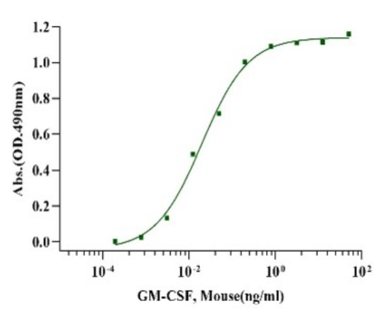
Purity determined by SDS-PAGE is greater than 95%, endotoxin is below 0.1 EU/μg, and the tag-free design avoids possible residual enzyme cleavage introduced by tag removal or spatial interference with the receptor binding interface. For bone marrow-derived dendritic cell induction, macrophage survival maintenance, and myeloid progenitor colony formation experiments, this configuration provides a reagent starting point with controllable background. Researchers should still establish dose-response curves in their own experimental systems to confirm that batch-specific activity falls within the expected range. After all, myeloid cells are extremely sensitive to microenvironmental signals, and fluctuations at the reagent level are often amplified by changes in cell state rather than buffered.
This article is reviewed and published by the technical expert team of UA
Disclaimer: This article partially utilizes artificial intelligence assistance in its creation. If any content involves copyright or intellectual property issues, please let us know and we promise to verify and remove it as soon as possible.
Purchase recombinant protein, choose 
UA protein focuses on providing various protein reagents, raw materials, and services required for drug research and development, cell therapy, gene therapy, and basic scientific research, including drug target proteins, immune checkpoint proteins, cytokines, tool enzymes, customized protein expression, and full-length transmembrane protein development. Nanjing Univ is committed to providing customers with high-quality products and professional services, and building a High-tech Biological Enterprise with International Competitiveness.
Target proteins | membrane proteins | cytokines | enzymes | viral antigens | protein customization
Nanjing UNIV Biotechnology Co., Ltd. Email:order@ua-bio.com Phone:+86-25-56221161

Product Information

Link:





©2021-2026 all rights reserved UNIV Biotechnology Co.,Ltd.
Privacy Policy
网站备案号 苏ICP备2025207586号-2 浙公网安备33010202004867号
 Business license (three certificates in one)
Tel:+86-25-56221161
E-mail:order@ua-bio.com
All products sold on this website shall not be used for clinical diagnosis or treatment of humans or animals, and shall only be used for non-medical purposes such as industrial or scientific research.


