




The Core Strategies and Experimental Protocols for In Vitro Activation and Expansion of Mouse T Cells
Author: Senior Product Manager of UA Views:32 


This article focuses on the core requirements of in vitro culture of mouse T cells, systematically elaborating the "three-signal mechanism" necessary for T cell activation and its molecular basis, while analyzing the technical principles and operational key points of solid-phase coating or magnetic bead methods based on anti-CD3/CD28 antibodies.
- Recent Advances
- Product Information
Recent Advances
Core Strategies and Experimental Protocols for In Vitro Activation and Expansion of Mouse T Cells
Summary
This article systematically elaborates on the "three-signal mechanism" required for T cell activation and its molecular basis, focusing on the core requirements for in vitro culture of mouse T cells. It analyzes the technical principles and operational details of solid-phase coating methods or magnetic bead-based methods using anti-CD3/CD28 antibodies.
This article systematically elaborates on the "three-signal mechanism" required for T cell activation and its molecular basis, focusing on the core requirements for in vitro culture of mouse T cells. It analyzes the technical principles and operational details of solid-phase coating methods or magnetic bead-based methods using anti-CD3/CD28 antibodies.
I. The "Three-Signal Mechanism" for Mouse T Cell Activation
Complete activation of T cells is a core step in initiating adaptive immune responses, relying on the coordinated input of three signals. The first signal (antigen-specific signal) is generated by the binding of the TCR-CD3 complex on the T cell surface to the MHC-antigen peptide complex on antigen-presenting cells (APCs), serving as the initiating signal for T cell activation. The second signal (co-stimulatory signal) is produced by the interaction between the CD28 molecule on T cells and the B7 ligands (CD80/CD86) on APCs, effectively overcoming T cell anergy and serving as the key to sustaining activation. The third signal (proliferation and survival signal) is mediated by the cytokine IL-2, which binds to the IL-2 receptor on T cells, activating the downstream JAK-STAT signaling pathway and driving T cells from the G0 phase into the cell cycle, enabling massive proliferation and maintaining cell viability and functional stability.
II. Technical Approaches to Simulate T Cell Activation In Vitro
In in vitro culture systems, anti-CD3/CD28 antibodies are standardized tools for mimicking APC function and providing T cell activation signals. Currently, two main technical approaches are widely used. The solid-phase antibody coating method immobilizes anti-CD3 and anti-CD28 antibodies directly on the culture plate surface, providing sustained and uniform stimulation, avoiding weak stimulation and uneven activation caused by free antibodies. The antibody-conjugated magnetic bead method utilizes three-dimensional magnetic beads to simulate the size and structure of APCs, forming efficient binding modes similar to immunological synapses with T cells. The superparamagnetic properties of the beads facilitate subsequent removal without interfering with downstream experiments. Both methods have their respective applications, and researchers can choose based on experimental needs.
III. Standardized Operational Procedures for In Vitro Activation and Expansion of Mouse T Cells
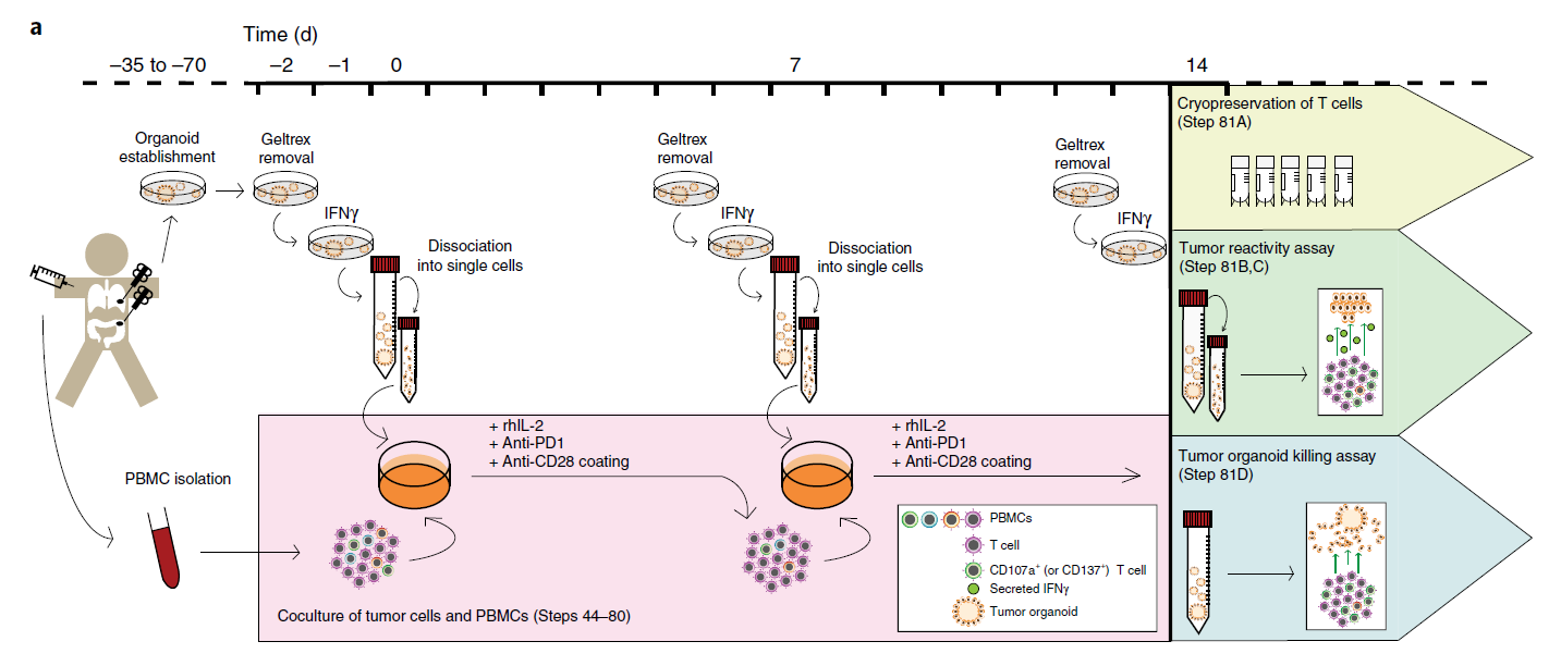
Taking the solid-phase coating method as an example, a typical procedure is as follows. Antibody coating stage: Dilute anti-mouse CD3 monoclonal antibody in PBS (final concentration: 10 µg/mL), add it to the culture plate, and incubate at 37°C for 2 hours or at 4°C overnight to ensure proper fixation. Cell seeding stage: After removing the coating solution, seed purified mouse T cells at an appropriate density (e.g., 8×10⁴ cells per well in a 96-well plate) in complete medium (RPMI-1640 + 10% FBS + β-mercaptoethanol + antibiotics). Activation and expansion stage: Add anti-CD28 antibody (final concentration: ~2 µg/mL) and recombinant mouse IL-2 (final concentration: 10 ng/mL) to the medium, and culture at 37°C with 5% CO₂. After 48-72 hours, T cell volume enlargement and morphological diversity can be observed. Fresh medium and IL-2 should be replenished as needed based on cell density.
IV. Conclusion
In vitro activation and expansion of mouse T cells are fundamental experimental steps in immunological function studies and preclinical cell therapy evaluations. The anti-CD3/CD28 antibody stimulation system, designed based on the "three-signal mechanism," combined with recombinant IL-2 for proliferation support, can efficiently simulate in vivo T cell activation processes in vitro. Standardized experimental procedures and high-quality reagent combinations enhance data reproducibility, providing reliable technical support for in-depth studies of T cell biology.
In the aforementioned study on in vitro activation and expansion of mouse T cells, standardized reagent combinations are crucial for ensuring experimental reproducibility. To meet this research need, Univ offers the Mouse T Cell Activation Kit/CellXViva Mouse T Cell Activation Kit, which includes anti-mouse CD3 monoclonal antibody, anti-mouse CD28 monoclonal antibody, and recombinant mouse IL-2 protein. All components have undergone rigorous concentration optimization and quality validation and can be directly used for in vitro activation and expansion experiments of T cells derived from mouse spleen, lymph nodes, or peripheral blood. This kit is suitable for basic immunology research, exploration of T cell signaling mechanisms, and construction of immune-related disease models.
This article is reviewed and published by the technical expert team of UA
Disclaimer: This article partially utilizes artificial intelligence assistance in its creation. If any content involves copyright or intellectual property issues, please let us know and we promise to verify and remove it as soon as possible.
Purchase recombinant protein, choose Nanjing UA-Bio
UA protein focuses on providing various protein reagents, raw materials, and services required for drug research and development, cell therapy, gene therapy, and basic scientific research, including drug target proteins, immune checkpoint proteins, cytokines, tool enzymes, customized protein expression, and full-length transmembrane protein development. Youai is committed to providing customers with high-quality products and professional services, and building a High-tech Biological Enterprise with International Competitiveness.
Target proteins | membrane proteins | cytokines | enzymes | viral antigens | protein customization
Nanjing UA Biotechnology Co., Ltd. Email:order@ua-bio.com Phone:+86-25-56221161

Product Information

Link:





©2021-2026 all rights reserved UA Biotechnology Co.,Ltd.
Privacy Policy 
网站备案号 浙ICP备2022019033号-2 浙公网安备33010202004867号
 Business license (three certificates in one)
 Tel:+86-25-56221161
E-mail:order@ua-bio.com
All products sold on this website shall not be used for clinical diagnosis or treatment of humans or animals, and shall only be used for non-medical purposes such as industrial or scientific research.
