




Technical Principles of pH-Sensitive IgG Labeling Reagents and Their Application in Antibody Internalization Research
Views:7 


This article focuses on the technical principles of pH-sensitive IgG fluorescent labeling reagents, systematically elucidating the molecular mechanism by which they utilize pH-dependent fluorescence signal changes to monitor antibody internalization processes, and analyzing their application value in antibody-drug conjugate screening and internalization kinetics research.
- Recent Advances
- Product Information
Recent Advances
Technical Principles of pH-Sensitive IgG Labeling Reagents and Their Applications in Antibody Internalization Research
Overview
This article systematically elaborates on the technical principles of pH-sensitive IgG fluorescent labeling reagents, focusing on their molecular mechanisms for monitoring antibody internalization processes through pH-dependent fluorescence signal changes. It also analyzes their application value in antibody-drug conjugate screening and internalization kinetics studies.
This article systematically elaborates on the technical principles of pH-sensitive IgG fluorescent labeling reagents, focusing on their molecular mechanisms for monitoring antibody internalization processes through pH-dependent fluorescence signal changes. It also analyzes their application value in antibody-drug conjugate screening and internalization kinetics studies.
I. The Critical Role of Antibody Internalization Activity Evaluation in ADC Drug Development
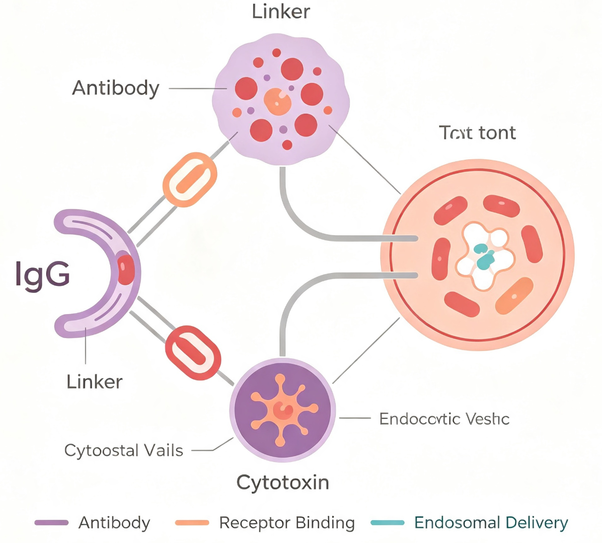
Antibody-drug conjugates (ADCs) consist of three components: a targeting antibody, a linker, and a small-molecule cytotoxic agent. Through the specific binding of the antibody to tumor cell surface antigens, the cytotoxic agent is delivered into target cells via receptor-mediated internalization pathways, achieving precise tumor cell killing. In this process, the efficiency of antibody internalization and its intracellular trafficking directly determine the therapeutic efficacy of ADCs. Therefore, accurately evaluating the internalization efficiency of candidate antibodies is a crucial step in the early stages of ADC drug development.
Traditional antibody labeling methods often struggle to distinguish between antibodies bound to the cell surface and those already internalized into the cells, leading to significant background interference in internalization activity assessments. The emergence of pH-sensitive fluorescent labeling technology provides an effective solution to this technical challenge.
II. Technical Principles of pH-Sensitive Fluorescent Labeling
The core design concept of pH-sensitive IgG labeling reagents lies in leveraging the optical properties of certain fluorophores that exhibit significant fluorescence intensity changes in acidic environments. Representative pH-sensitive labeling reagents, such as Max(Green), demonstrate markedly enhanced fluorescence intensity at pH 5.0 (acidic conditions) and significantly reduced fluorescence at pH 7.4 (neutral conditions). This property aligns perfectly with cellular internalization processes—when the antibody-pH-sensitive labeling reagent complex binds to the cell surface (neutral pH environment), the fluorescence signal remains low; however, when the complex is internalized into endosomes or lysosomes (acidic microenvironment, pH 4.5-5.5), the fluorescence signal increases substantially.
Through this mechanism, pH-sensitive labeling reagents can effectively distinguish between cell surface-bound antibodies and internalized antibodies, significantly reducing background interference and enabling precise quantification of antibody internalization activity.
III. Molecular Design and Operational Workflow of IgG Labeling Reagents
Max(Green) pH-sensitive IgG labeling reagent is a fluorescent dye-labeled Fc-binding protein with a molecular weight of approximately 31-33 kDa. This reagent can specifically bind to IgG antibodies from various species (including human IgG1, IgG2, IgG3, IgG4; rabbit IgG; mouse IgG1, IgG2a, IgG2b, and IgG3, etc.), forming fluorescently labeled antibody-reagent complexes.
The typical workflow includes the following steps: First, the test antibody is mixed with the pH-sensitive labeling reagent at the recommended ratio and incubated at room temperature in the dark for a specified duration to form antibody-reagent complexes. Next, these complexes are co-incubated with target cells at 37°C to allow antibody internalization. Subsequently, fluorescence intensity (detected using FITC or AF488 channels) is measured by flow cytometry, and with appropriate controls, the antibody internalization efficiency can be quantitatively evaluated. The entire labeling process is straightforward, requires no complex purification steps, and is compatible with high-throughput screening modes.
IV. Conclusion
pH-sensitive IgG labeling technology leverages its intelligent optical response to acidic microenvironments, effectively addressing the technical challenge of distinguishing between surface-bound and internalized antibodies with traditional labeling methods. It provides a precise and efficient analytical tool for ADC drug development and antibody internalization mechanism research. The pH-sensitive IgG labeling reagent Max(Green), with its broad compatibility, simple workflow, and excellent pH-responsive performance, holds significant application value in antibody drug discovery and fundamental cell biology research.
The pH-sensitive IgG labeling reagent Max(Green) developed by Univ is specifically designed for antibody internalization studies. Its key features include: Based on MaxGreen fluorescent dye, with excitation/emission wavelengths of 488/520 nm, highly compatible with flow cytometry FITC or AF488 detection channels; broad-spectrum recognition of IgG antibodies from multiple species and subtypes; provided in lyophilized form, stable for 12 months at -20 to -80°C, and can be stored for 1-2 weeks at 2-8°C in sterile conditions after reconstitution; optimized for high labeling efficiency and ease of operation.
Disclaimer: This article partially utilizes artificial intelligence assistance in its creation. If any content involves copyright or intellectual property issues, please let us know and we promise to verify and remove it as soon as possible.
Purchase recombinant protein, choose Nanjing UA-Bio
UA protein focuses on providing various protein reagents, raw materials, and services required for drug research and development, cell therapy, gene therapy, and basic scientific research, including drug target proteins, immune checkpoint proteins, cytokines, tool enzymes, customized protein expression, and full-length transmembrane protein development. Youai is committed to providing customers with high-quality products and professional services, and building a High-tech Biological Enterprise with International Competitiveness.
Target proteins | membrane proteins | cytokines | enzymes | viral antigens | protein customization
Nanjing UA Biotechnology Co., Ltd. Email:order@ua-bio.com Phone:+86-25-56221161

Product Information

Link:





©2021-2026 all rights reserved UA Biotechnology Co.,Ltd.
Privacy Policy 
网站备案号 浙ICP备2022019033号-2 浙公网安备33010202004867号
 Business license (three certificates in one)
 Tel:+86-25-56221161
E-mail:order@ua-bio.com
All products sold on this website shall not be used for clinical diagnosis or treatment of humans or animals, and shall only be used for non-medical purposes such as industrial or scientific research.
