




Technical Pathway and High-Throughput Applications of Fluorescence-Based Cell Viability Assays
Views:6 


This article focuses on fluorescence signal-based cell viability detection technologies, systematically elaborating on the molecular principles and operational characteristics of two technical approaches targeting live cell protease activity and cell membrane integrity as primary detection markers. It analyzes the application advantages of this technology in drug screening and cellular function research.
- Recent Advances
- Product Information
Recent Advances
Technical Pathways and High-Throughput Applications of Fluorescence-Based Cell Viability Assays
Overview
This article systematically elaborates on fluorescence-based cell viability detection technologies, focusing on two technical pathways targeting intracellular protease activity and cell membrane integrity. It analyzes the molecular principles, operational characteristics, and application advantages in drug screening and cellular function research.
This article systematically elaborates on fluorescence-based cell viability detection technologies, focusing on two technical pathways targeting intracellular protease activity and cell membrane integrity. It analyzes the molecular principles, operational characteristics, and application advantages in drug screening and cellular function research.
I. Cell Viability Detection: Evolution from Traditional Methods to Fluorescence Technologies
Cell viability assays are among the most frequently conducted experiments in cell biology, pharmacology, and cancer research. Whether evaluating the cytotoxicity of candidate compounds, assessing the effects of cytokines on cell proliferation, or optimizing cell culture conditions, an accurate, simple, and reproducible viability detection method is essential. Traditional cell viability detection methods primarily include dye exclusion (e.g., trypan blue staining), tetrazolium salt reduction (e.g., MTT assay), and ATP bioluminescence. While these methods have their respective applications, they also suffer from limitations such as cumbersome procedures, endpoint detection, or limited sensitivity.
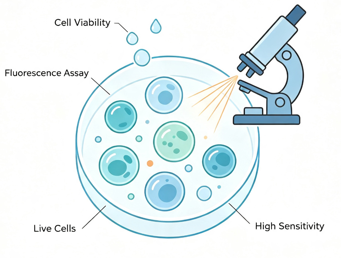
With the widespread adoption of fluorescent dyes and detection instruments, fluorescence-based cell viability assays are becoming increasingly popular in drug screening and basic research due to their high sensitivity, broad linear range, and homogeneous operation.
II. Fluorescence Detection Strategy Based on Intracellular Protease Activity
Intracellular constitutive protease activity is a robust and reliable indicator for assessing the number of viable cells. In living cells, specific proteases are continuously expressed and maintain activity, which is crucial for cell survival. Detection methods based on this principle employ fluorescent substrates (e.g., peptides containing AFC or AMC groups) that can penetrate intact cell membranes. These substrates freely enter live cells and are specifically recognized and cleaved by active intracellular proteases, releasing free fluorescent groups. Since dead cells lose membrane integrity and their internal proteases rapidly deactivate, they cannot generate signals, resulting in a strong linear relationship between detected fluorescence intensity and the number of viable cells.
The core advantage of this technical pathway lies in its homogeneous, non-lytic operation mode. The experimental procedure consists of only three steps: "add reagent-incubate-read," eliminating the need for washing or cell lysis and minimizing operational errors. Moreover, this detection method is reversible and suitable for continuous monitoring, making it ideal for time-course experiments or as a normalization parameter in multiplex assays.
III. Live/Dead Cell Dual-Staining Strategy Based on Cell Membrane Integrity
The live/dead cell dual-staining strategy based on cell membrane integrity is the most widely used viability detection method in flow cytometry and fluorescence microscopy. A representative combination is the dual-staining system of Calcein-AM and propidium iodide (PI). Calcein-AM is a non-fluorescent molecule that penetrates intact cell membranes and is hydrolyzed by intracellular nonspecific esterases in live cells, generating negatively charged green fluorescent Calcein molecules that are trapped in the cytoplasm due to their inability to freely cross cell membranes. PI, on the other hand, can only penetrate dead cells with compromised membrane integrity, binding to DNA and emitting red fluorescence. By simultaneously detecting green and red fluorescence using a fluorescence microscope or flow cytometer, viable and dead cell populations can be visually distinguished. This technique is widely used in cytotoxicity assessment, immune cell function analysis, and quality control in organoid culture.
IV. Application Advantages of Fluorescence Detection in Drug Screening and Functional Research
In high-throughput drug screening systems, fluorescence-based cell viability assays demonstrate unique advantages. Their high sensitivity ensures good linearity of detection signals even with low cell numbers, saving both cells and compounds. Fluorescence detection exhibits strong tolerance to common interferents such as phenol red and serum in culture media, resulting in superior data stability. The homogeneous operation is highly compatible with automated liquid handling workstations, making it suitable for large-scale screening. Additionally, fluorescence-based detection systems can be flexibly combined with various downstream analysis platforms, facilitating standardized correction of experimental results using cell viability data.
V. Conclusion
Fluorescence-based cell viability detection technologies, with their high sensitivity, simple operation, and excellent platform compatibility, have become indispensable tools in modern biomedical research. Whether using homogeneous detection methods based on protease activity or dual-staining methods based on membrane integrity, these techniques offer unique value in their respective application scenarios.
To address the aforementioned cell viability detection needs, UniLove offers the UA-Glo® Fluorescent Cell Viability Assay. This kit employs an optimized homogeneous fluorescence detection system based on the principle of live cell-specific protease activity, eliminating the need for cell lysis or washing steps. It is compatible with both adherent and suspension cells, supports 96-well and 384-well plate formats, and is suitable for high-throughput operations. The assay features a simple protocol, reducing overall processing time, and provides stable signals with flexible reading time windows. This kit is ideal for compound cytotoxicity screening, cell proliferation and survival analysis, antibody drug cell-killing activity evaluation, and viability monitoring in organoid and primary cell cultures.
Disclaimer: This article partially utilizes artificial intelligence assistance in its creation. If any content involves copyright or intellectual property issues, please let us know and we promise to verify and remove it as soon as possible.
Purchase recombinant protein, choose Nanjing UA-Bio
UA protein focuses on providing various protein reagents, raw materials, and services required for drug research and development, cell therapy, gene therapy, and basic scientific research, including drug target proteins, immune checkpoint proteins, cytokines, tool enzymes, customized protein expression, and full-length transmembrane protein development. Youai is committed to providing customers with high-quality products and professional services, and building a High-tech Biological Enterprise with International Competitiveness.
Target proteins | membrane proteins | cytokines | enzymes | viral antigens | protein customization
Nanjing UA Biotechnology Co., Ltd. Email:order@ua-bio.com Phone:+86-25-56221161

Product Information

Link:





©2021-2026 all rights reserved UA Biotechnology Co.,Ltd.
Privacy Policy 
网站备案号 浙ICP备2022019033号-2 浙公网安备33010202004867号
 Business license (three certificates in one)
 Tel:+86-25-56221161
E-mail:order@ua-bio.com
All products sold on this website shall not be used for clinical diagnosis or treatment of humans or animals, and shall only be used for non-medical purposes such as industrial or scientific research.
