




Fluorescent Cell Viability Detection Technology: Principles, Advantages, and Applications
Views:5 


This article focuses on the technical principles of fluorescence-based cell viability detection, systematically elaborating on the molecular basis of two core detection strategies: live-cell protease activity and cell membrane integrity, and analyzing their technical advantages compared to traditional colorimetric methods.
- Recent Advances
- Product Information
Recent Advances
Fluorescent Cell Viability Detection Technology: Principles, Advantages, and Applications
Overview
This article focuses on the technical principles of fluorescent cell viability detection, systematically elaborating on the molecular basis of two core detection strategies—based on live-cell protease activity and cell membrane integrity—and analyzing their advantages over traditional colorimetric methods.
This article focuses on the technical principles of fluorescent cell viability detection, systematically elaborating on the molecular basis of two core detection strategies—based on live-cell protease activity and cell membrane integrity—and analyzing their advantages over traditional colorimetric methods.
I. The Central Role of Cell Viability Detection in Life Science Research.
Accurate and rapid determination of cell viability and cell count is a critical prerequisite and foundation for cell biology experiments. Whether it's cytotoxicity assessment in drug screening, drug sensitivity analysis in tumor research, or cell quality control in vaccine development, cell viability detection is an indispensable core component. Traditional cell viability detection methods mainly fall into two categories: Trypan blue staining, which selectively stains dead cells for manual counting, is labor-intensive and subjective; MTT colorimetric assays, based on mitochondrial dehydrogenase activity reducing tetrazolium salts to produce colored products, are cost-effective but suffer from interference by cell metabolic states, require dissolution steps, and are unsuitable for suspension cells.
With the maturation of fluorescent detection technology, fluorescent-based cell viability detection methods have become the mainstream solution in the field due to their high sensitivity, ease of operation, and objective results.
II. Core Technical Principles of Fluorescent Cell Viability Detection.
Currently, mainstream fluorescent cell viability detection methods are primarily based on two biological strategies.
One important technical approach is the detection strategy based on live-cell protease activity. Its principle relies on measuring the activity of a conserved and constitutively expressed protease in live cells. This method uses a cell-permeable fluorescent peptide substrate (e.g., Gly-Phe-AFC). The substrate freely enters intact live cells, where it is cleaved by live-cell proteases to release a fluorescent group, generating a signal proportional to the number of live cells. When cell membrane integrity is lost, the protease leaks into the extracellular medium and is deactivated, ensuring high specificity for live cells. This method is non-lytic and homogeneous, requiring only three steps—"add-incubate-read"—making it particularly suitable for continuous monitoring or subsequent multiplex assays.
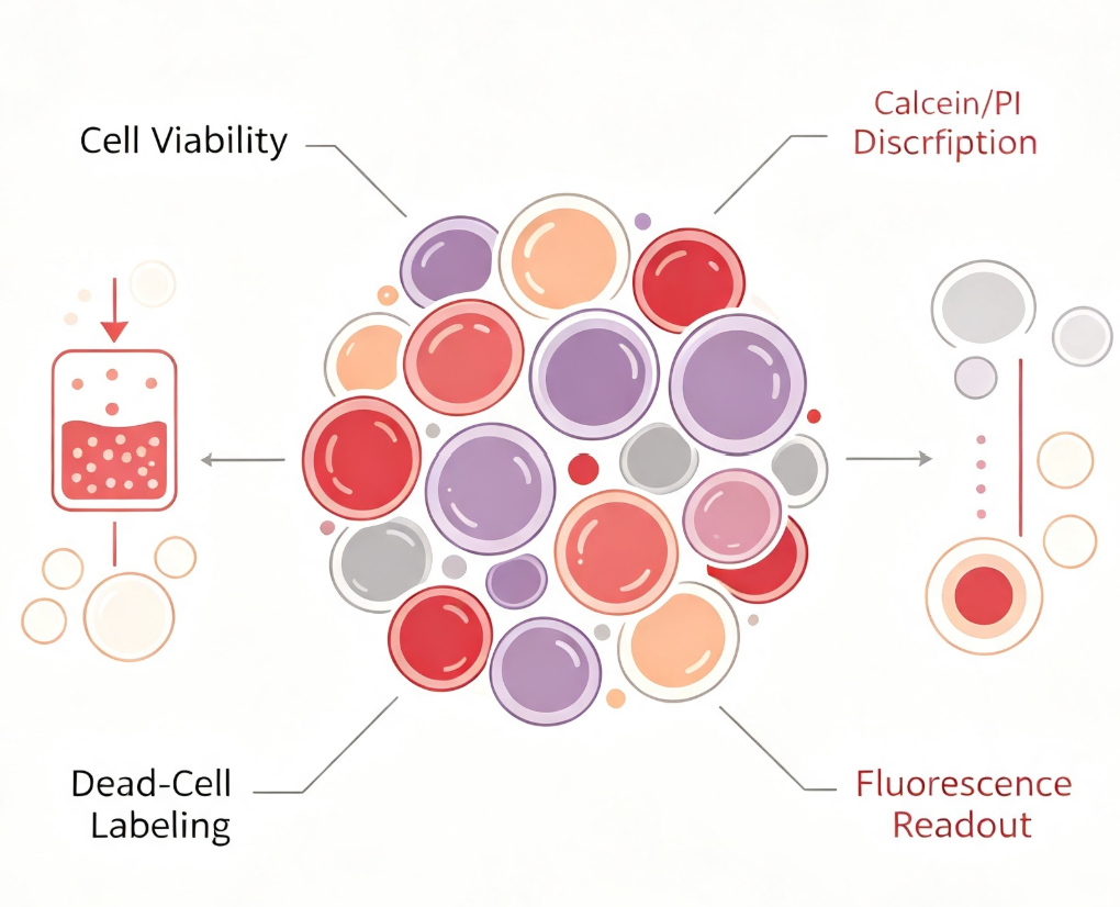
Another widely used method is the dual-staining strategy for live/dead cells based on cell membrane integrity, exemplified by the Calcein-AM/PI dual-staining assay. Calcein-AM is a non-fluorescent molecule that penetrates intact cell membranes and is hydrolyzed by intracellular esterases into highly green-fluorescent Calcein (Ex/Em: 495/515 nm), labeling live cells. Propidium iodide (PI), however, cannot penetrate intact cell membranes and only enters dead cells with compromised membranes, binding to nucleic acids and emitting red fluorescence (Ex/Em: 535/617 nm). This method allows for visual differentiation of live and dead cells via fluorescence microscopy or flow cytometry and is widely used in cytotoxicity assessment and cell quality control.
III. Technical Advantages of Fluorescent Detection Methods.
Compared to traditional colorimetric methods, fluorescent detection methods exhibit several significant technical advantages in cell viability analysis. First, fluorescent signals offer higher sensitivity and signal-to-noise ratios, enabling detection of smaller numbers of live cells or more subtle viability changes. Second, fluorescent methods typically require no cell lysis or medium removal, making them simpler and faster to perform, with compatibility for both adherent and suspension cells. Third, fluorescent methods are less affected by medium components (e.g., phenol red, serum) and reaction times, yielding more stable and reliable results. Additionally, some fluorescent reagents have low cytotoxicity, allowing for continuous monitoring of the same culture well.
IV. Conclusion.
Fluorescent cell viability detection technology, with its high sensitivity, ease of operation, and excellent compatibility, has become a fundamental tool in modern cell biology research and drug development. The different detection strategies—based on live-cell protease activity or cell membrane integrity—provide researchers with flexible options. The UA-Glo® Fluorescent Cell Viability Assay Kit, as a leading product in this field, offers reliable technical support for various experimental scenarios involving cell viability.
To address the above cell viability detection needs, U-A Biotech offers the UA-Glo® Fluorescent Cell Viability Assay. This kit features an optimized fluorescent detection system with the following key characteristics: homogeneous single-reagent operation, no washing or cell lysis steps required, compatibility with high-throughput detection modes; flexible integration with various downstream analysis systems for standardized correction of experimental results using cell viability data; suitability for drug cytotoxicity assessment, cell proliferation analysis, and high-throughput compound screening, among other research applications.
Disclaimer: This article partially utilizes artificial intelligence assistance in its creation. If any content involves copyright or intellectual property issues, please let us know and we promise to verify and remove it as soon as possible.
Purchase recombinant protein, choose Nanjing UA-Bio
UA protein focuses on providing various protein reagents, raw materials, and services required for drug research and development, cell therapy, gene therapy, and basic scientific research, including drug target proteins, immune checkpoint proteins, cytokines, tool enzymes, customized protein expression, and full-length transmembrane protein development. Youai is committed to providing customers with high-quality products and professional services, and building a High-tech Biological Enterprise with International Competitiveness.
Target proteins | membrane proteins | cytokines | enzymes | viral antigens | protein customization
Nanjing UA Biotechnology Co., Ltd. Email:order@ua-bio.com Phone:+86-25-56221161

Product Information

Link:





©2021-2026 all rights reserved UA Biotechnology Co.,Ltd.
Privacy Policy 
网站备案号 浙ICP备2022019033号-2 浙公网安备33010202004867号
 Business license (three certificates in one)
 Tel:+86-25-56221161
E-mail:order@ua-bio.com
All products sold on this website shall not be used for clinical diagnosis or treatment of humans or animals, and shall only be used for non-medical purposes such as industrial or scientific research.
