




Mouse-derived CD3E/CD3G heterodimer: A core tool for T-cell bispecific antibody development and a key to affinity modulation
Views:10 


This article focuses on the central role of the CD3 antigen in the development of bispecific antibody drugs, systematically elaborating on the structural basis and physiological necessity of the heterodimer formation between CD3E and CD3G, and analyzing the critical role of CD3 antibody affinity in balancing efficacy and safety.
- Recent Advances
- Product Information
Recent Advances
Murine CD3E/CD3G Heterodimer: A Core Tool for T-cell Bispecific Antibody Development and the Key to Affinity Modulation
Summary
This article focuses on the central role of CD3 antigens in the development of bispecific antibody drugs, systematically elaborating on the structural basis and physiological necessity of CD3E and CD3G forming heterodimers, and analyzing the critical role of CD3 antibody affinity in balancing efficacy and safety.
This article focuses on the central role of CD3 antigens in the development of bispecific antibody drugs, systematically elaborating on the structural basis and physiological necessity of CD3E and CD3G forming heterodimers, and analyzing the critical role of CD3 antibody affinity in balancing efficacy and safety.
I. The Development Background of CD3 Bispecific Antibodies and the Principle of Affinity Balance.
With the rapid development of bispecific/multispecific antibodies and tumor immunotherapies, CD3 antibodies have once again become a focal point in the field of antibody drug development. According to statistics, nearly half of the bispecific antibodies in clinical stages target CD3 antigens as one of their binding sites. Bispecific antibodies recruit T cells to tumor sites and activate their cytotoxic functions by binding to tumor-associated antigens on one end and CD3 on T cells on the other, achieving precise tumor clearance.
However, the development of CD3 bispecific antibodies faces a core "affinity dilemma"—the affinity of the CD3 antibody end for CD3 antigens must be precisely regulated within a specific range. Literature and industry consensus indicate that if the affinity is too strong (e.g., below 1 nM), it may excessively activate T cells, triggering severe safety risks such as cytokine release syndrome (CRS). Conversely, if the affinity is too weak (e.g., above 1 μM), it cannot effectively recruit and activate T cells at tumor sites, resulting in insufficient efficacy. Studies show that when the affinity of the CD3 antibody end for CD3 antigens falls within the range of 10 nM to 100 nM, an optimal therapeutic window and in vivo efficacy can typically be achieved.
As the development paradigm shifts from "pursuing the strongest binding" to "finding the optimal affinity"—a principle that also applies to immune agonist antibodies (e.g., anti-CD137) or safer CAR-T designs—the accurate quantification of antibody-antigen affinity has become a key attribute parameter determining drug safety and efficacy.
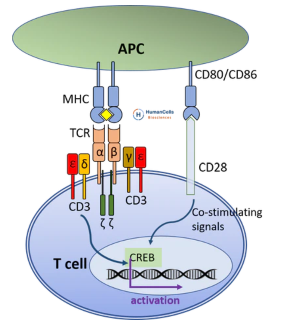
II. The Molecular Structure of CD3 Antigens and the Physiological Necessity of Heterodimer Conformation.
CD3 is a group of type I transmembrane proteins found on the surface of T cells, comprising four subtypes: CD3D (CD3δ), CD3E (CD3ε), CD3G (CD3γ), and CD3Z (CD3ζ). In the TCR-CD3 complex, CD3D and CD3E form a heterodimer, while CD3G and CD3E form another heterodimer. These heterodimers preferentially associate with the TCR α and β chains, respectively, forming a stable octameric structure.
When antibody drug development involves targeting CD3, it strictly refers to targeting human CD3E antigen (UniProt ID: P07766). However, the biological characteristics of CD3E dictate that it cannot be regarded as an independent functional unit—under physiological conditions, CD3 antigens on T cells exist as CD3D/CD3E and CD3G/CD3E heterodimers. Due to protein flexibility, the spatial conformation of CD3E in its monomeric state differs significantly from that in its heterodimerized state, which directly affects the recognition and binding efficiency of therapeutic antibodies to epitopes.
III. Conformation-Dependent Insights Revealed by Activity Validation: Differential Recognition of CD3 Heterodimers by Clinical Antibodies.
The biological importance of CD3 antigen heterodimer conformations can be validated through binding data from clinical-stage antibodies. For example, the anti-CD3 antibody Foralumab exhibits an affinity of approximately 8 nM for the CD3E/CD3G heterodimer, while its affinity for the CD3E/CD3D heterodimer is about 0.1 nM—a difference of nearly two orders of magnitude. This disparity indicates that the epitope conformation of CD3E undergoes significant changes when forming heterodimers with different partners, particularly in the spatial structure of the antibody-binding region. These findings underscore the necessity of using heterodimeric proteins with native conformations for antibody screening and evaluation.
IV. Conclusion.
CD3E and CD3G form the core signaling module of the TCR-CD3 complex through heterodimerization. Precise modulation of CD3 antibody affinity within the 10-100 nM range is key to balancing bispecific antibody efficacy and CRS risk. Since CD3E exhibits different epitope conformations when forming heterodimers with different partners, and its spatial structure differs between monomeric and heterodimeric states, it is essential to use correctly conformed heterodimeric recombinant proteins for antibody screening and evaluation. The murine CD3E&CD3G heterodimer protein provided by U-Trust, with its precise molecular design, native conformation ensured by the HEK293 expression system, and high antibody-binding activity validated by ELISA, offers a reliable tool for screening bispecific antibody candidates and preclinical evaluation.
To address these research needs, U-Trust offers CD3E&CD3G Heterodimer, Fc, His Tag&Fc, Flag Tag Protein, Mouse. This product is prepared using a HEK293 co-expression system and includes the extracellular domains of murine CD3E (UniProt NP_031674.1, amino acids D23-D108) and CD3G (UniProt NP_033980.1, amino acids Q23-S116), each fused with different Fc tags (His and Flag). Two-step affinity chromatography ensures high purity (>95%) and equimolar expression of the heterodimer.
Disclaimer: This article partially utilizes artificial intelligence assistance in its creation. If any content involves copyright or intellectual property issues, please let us know and we promise to verify and remove it as soon as possible.
Purchase recombinant protein, choose Nanjing UA-Bio
UA protein focuses on providing various protein reagents, raw materials, and services required for drug research and development, cell therapy, gene therapy, and basic scientific research, including drug target proteins, immune checkpoint proteins, cytokines, tool enzymes, customized protein expression, and full-length transmembrane protein development. Youai is committed to providing customers with high-quality products and professional services, and building a High-tech Biological Enterprise with International Competitiveness.
Target proteins | membrane proteins | cytokines | enzymes | viral antigens | protein customization
Nanjing UA Biotechnology Co., Ltd. Email:order@ua-bio.com Phone:+86-25-56221161

Product Information

Link:





©2021-2026 all rights reserved UA Biotechnology Co.,Ltd.
Privacy Policy 
网站备案号 浙ICP备2022019033号-2 浙公网安备33010202004867号
 Business license (three certificates in one)
 Tel:+86-25-56221161
E-mail:order@ua-bio.com
All products sold on this website shall not be used for clinical diagnosis or treatment of humans or animals, and shall only be used for non-medical purposes such as industrial or scientific research.
