

A Systematic Elucidation of MOG Antibody-Associated Disease: Pathogenesis, Clinical Features, and Treatment Strategies
Views:13 


This article systematically elaborates on the structural characteristics and immunogenicity of the myelin oligodendrocyte glycoprotein (MOG), the pathogenic mechanisms of MOG antibodies, and the historical evolution of detection methods for MOG antibody-associated disease (MOG-AD). It analyzes the diverse clinical phenotypes and imaging features of the disease across different age groups and discusses its treatment principles and prognosis.
- Recent Advances
- Product Information
Recent Advances
MOG Antibody-Associated Disease: A Systematic Review of Pathogenesis, Clinical Features, and Treatment Strategies
Summary: This article systematically reviews the structural characteristics and immunogenicity of myelin oligodendrocyte glycoprotein (MOG), the pathogenic mechanisms of MOG antibodies, the historical evolution of detection methods, the diverse clinical phenotypes and imaging features across different age groups, and discusses treatment principles and prognosis.
1. Structural Characteristics and Biological Significance of MOG
Myelin oligodendrocyte glycoprotein is a type I transmembrane glycoprotein composed of 218 amino acids, specifically expressed on the outermost myelin membrane of oligodendrocytes in the central nervous system (CNS) and belonging to the immunoglobulin superfamily. Among all myelin protein components, MOG has an extremely low abundance, accounting for less than 0.05% of total myelin proteins. Despite its scarcity, its unique localization—exposed on the outermost layer of myelin and facing the extracellular space—makes it a critical target for immune recognition.
The physiological functions of MOG remain incompletely understood, but based on its molecular structure, it is speculated to play roles in maintaining myelin structural stability, regulating oligodendrocyte cytoskeleton, and participating in immune modulation through complement activation. Importantly, MOG is recognized as a key autoantigen in the CNS. Animal studies have confirmed that MOG antibodies are pathogenic in experimental autoimmune encephalomyelitis models, exacerbating demyelination and inflammatory responses.
2. Evolution of MOG Antibody Detection Methods and Establishment of Diagnostic Criteria
Early studies on MOG antibodies primarily used the extracellular domain of MOG synthesized in E. coli as the antigen, detected via enzyme-linked immunosorbent assay (ELISA) or immunoblotting. However, these methods could only identify linear epitopes of the MOG extracellular domain, and such antibodies could be detected in both healthy individuals and multiple sclerosis (MS) patients, lacking disease specificity. Animal studies have shown that only antibodies targeting the correct three-dimensional conformation of MOG are pathogenic.
This understanding led to a fundamental shift in detection technology. Cell-based assays (CBA) emerged, which involve transfecting RNA encoding full-length human MOG into engineered cells to express MOG protein with its native conformation on the cell membrane. Serum antibodies targeting conformational epitopes are then detected via cell-based immunofluorescence. CBA significantly improves specificity and clinical relevance. International consensus now recommends CBA as the standard method for MOG antibody detection and emphasizes that the diagnosis of MOG-AD requires both antibody positivity and typical clinical manifestations, with antibody positivity being a prerequisite for diagnosis.
3. Pathological Features and Pathogenesis of MOG-AD
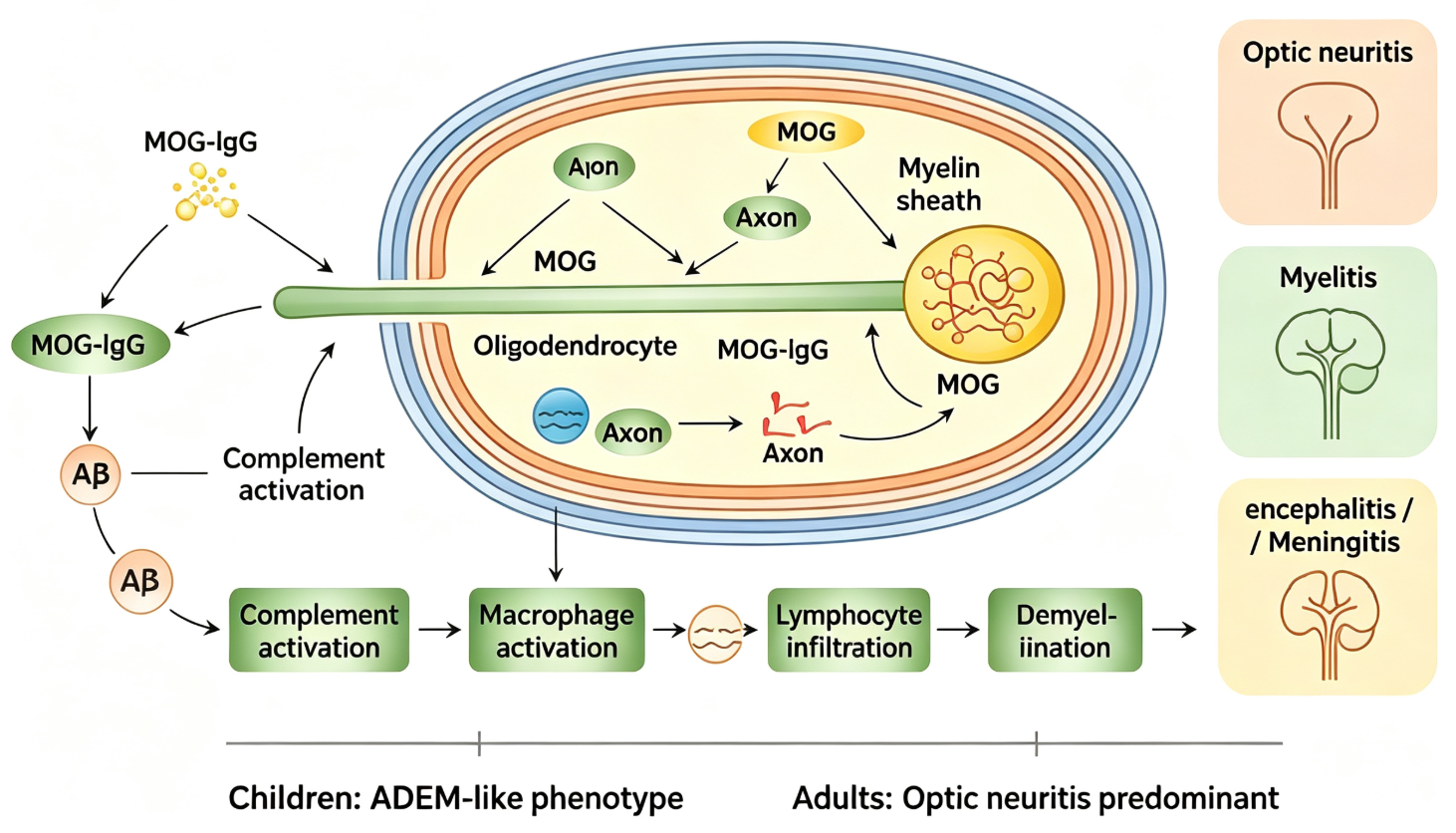
Due to limited case numbers, current understanding of MOG-AD pathology primarily comes from brain biopsy materials. Its basic pathological changes resemble other inflammatory demyelinating diseases, characterized by perivascular lymphocyte infiltration, demyelination, deposition of IgG and complement components, and macrophage activation. A few cases show granuloma-like structures around blood vessels, with lesions affecting both brain parenchyma and leptomeninges. These findings suggest MOG-AD is a humoral immune-mediated injury involving antibodies and complement.
The production of pathogenic MOG antibodies is thought to begin with transient increases in blood-brain barrier permeability—when CNS infections or other events occur, MOG antigens may leak into peripheral circulation and be recognized by the immune system. Animal studies further reveal that gut microbiota can assist in activating MOG-specific CD4+ T cells and B cells, promoting MOG antibody production. When the blood-brain barrier is compromised again, peripherally generated pathogenic antibodies enter the CNS, attacking MOG-expressing myelin via complement-dependent cytotoxicity and antibody-dependent cellular cytotoxicity. Inflammatory responses at lesion sites further recruit lymphocytes and macrophages, amplifying cascades through cytokines like IL-12, IL-17, and TNF-α, leading to demyelinating damage.
4. Clinical Phenotypes of MOG-AD and Their Age-Related Patterns
MOG-AD exhibits marked heterogeneity and age-related clinical manifestations. The MOG antibody positivity rate is significantly higher in children (~40%) than in adults (~22%), with a slight female predominance and no clear racial clustering. Clinically, children predominantly present with acute disseminated encephalomyelitis-like symptoms, while adults most commonly exhibit optic neuritis (ON). Other phenotypes include myelitis, brainstem encephalitis, and meningitis.
Optic neuritis is the most common phenotype, characterized by rapid vision loss, often accompanied by eye pain and pain with eye movement. Unlike ON in MS or aquaporin-4 (AQP4) antibody-positive neuromyelitis optica spectrum disorder (NMOSD), MOG-AD-related ON has the following features: high bilateral involvement (>50%), prominent optic disc edema, predominant anterior optic nerve involvement (retrobulbar and intraorbital segments), and rare chiasmal involvement. Imaging shows optic nerve thickening and swelling, with enhancement extending to intraorbital soft tissues on contrast scans—a relatively specific feature of MOG-ON. Despite severe visual impairment, most patients respond well to immunotherapy, with a much lower blindness rate than AQP4-IgG-positive NMOSD.
Myelitis accounts for ~20% of MOG-AD cases, most commonly affecting the cervical and thoracic spine, with high conus medullaris involvement and a high incidence of erectile dysfunction in male patients. Lesions often extend over three vertebral segments but can also present as short-segment or multifocal non-contiguous lesions. MRI typically shows lesions confined to gray matter, appearing as "H"-shaped hyperintensity on axial views and linear hyperintensity along the anterior spinal cord on sagittal views, aiding differentiation from NMOSD and MS.
Encephalitis and meningitis present diversely, including psychiatric symptoms, seizures, and altered consciousness. ~20% of patients exhibit meningitis symptoms with elevated intracranial pressure and CSF cell counts. MRI commonly shows subcortical and deep nuclear T2 hyperintensity, sometimes with leptomeningeal enhancement, frequently involving infratentorial structures like the thalamus, brainstem, and cerebellum.
Rare phenotypes include tumor-like demyelinating lesions (mimicking neoplasms on imaging) and pontine linear enhancement (resembling CLIPPERS syndrome), further complicating diagnosis.
6. Conclusion
MOG-AD is an independent CNS inflammatory demyelinating disease mediated by MOG antibodies, distinct from multiple sclerosis and AQP4-IgG-positive NMOSD in pathogenesis, clinical manifestations, imaging features, treatment response, and prognosis. CBA detection of conformation-specific MOG antibodies is the cornerstone of diagnosis. As understanding of the disease deepens, its clinical phenotype spectrum continues to expand, and standardized treatment regimens require further prospective studies. High-quality recombinant MOG protein supply provides a critical foundation for basic research and diagnostic optimization. Univ offers MOG His Tag Protein, Human, suitable for establishing and validating MOG antibody detection methods, quality control in serological screening of MOG-AD patients, and basic research on MOG antigen structure and immunogenicity.
Disclaimer: This article partially utilizes artificial intelligence assistance in its creation. If any content involves copyright or intellectual property issues, please let us know and we promise to verify and remove it as soon as possible.
Purchase recombinant protein, choose Nanjing UA-Bio
UA protein focuses on providing various protein reagents, raw materials, and services required for drug research and development, cell therapy, gene therapy, and basic scientific research, including drug target proteins, immune checkpoint proteins, cytokines, tool enzymes, customized protein expression, and full-length transmembrane protein development. Youai is committed to providing customers with high-quality products and professional services, and building a high-tech enterprise with international competitiveness.
Target proteins | membrane proteins | cytokines | enzymes | viral antigens | protein customization
Nanjing UA Biotechnology Co., Ltd. Email:order@ua-bio.com Phone:0571-87565022

Product Information

Link:





©2021-2026 all rights reserved UA Biotechnology Co.,Ltd.

网站备案号 浙ICP备2022019033号-2 浙公网安备33010202004867号
 Business license (three certificates in one)
 Tel:+86-0571-87565022
E-mail:order@ua-bio.com
All products sold on this website shall not be used for clinical diagnosis or treatment of humans or animals, and shall only be used for non-medical purposes such as industrial or scientific research.
