

Standardized construction protocol and culture system for normal mouse lung organoids
Views:15 


This article focuses on the in vitro construction and cultivation of mouse lung organoids, systematically detailing the standardized operational procedures from tissue isolation, enzymatic digestion, Matrigel embedding to primary culture. It analyzes key technical points in critical steps such as passaging and expansion, cryopreservation and revival, and explores how to stably obtain lung organoid models with typical cavity structures through precise control of operational steps.
- Recent Advances
- Product Information
Recent Advances
Standardized Construction Protocol and Culture System for Mouse Normal Lung Organoids
Brief Introduction: This article systematically elaborates on the standardized operational procedures for the in vitro construction and culture of mouse lung organoids, from tissue isolation, enzymatic digestion, Matrigel embedding to primary culture. It analyzes the technical key points of critical steps such as passaging expansion, cryopreservation, and recovery, and discusses how to stably obtain lung organoid models with typical cavity structures through precise control of operational steps.
1. Research Value and Experimental Preparation of Organoid Models
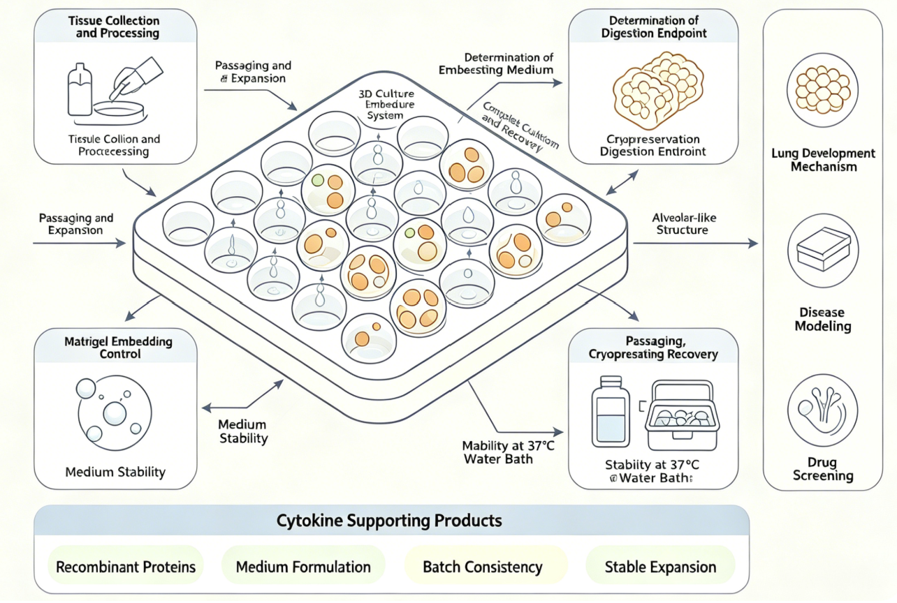
The mouse lung organoid model is a rapidly developing three-dimensional in vitro culture system in recent years. It is formed by the self-organization of adult stem cells and progenitor cells from lung tissue under the support of Matrigel, capable of highly simulating the three-dimensional structure and cellular diversity of in vivo lung tissue. Compared to traditional two-dimensional cell culture, organoids are closer to the physiological state and have unique application advantages in areas such as lung development mechanisms, disease modeling, and drug screening. However, organoid construction is extremely sensitive to operational details, from sample processing to medium selection, each step is crucial to the success of the experiment. Therefore, establishing a standardized operational procedure is essential to reduce technical barriers and improve experimental reproducibility.
Before starting the experiment, systematic preparation of reagents and consumables is required. Core reagents include Matrigel, tissue digestion solution, rinsing solution, DPBS, red blood cell lysis buffer, organoid digestion solution, cryopreservation solution, and complete medium. All consumables that come into contact with cells should be pre-treated with rinsing solution to prevent cell adhesion loss.
2. Collection and Cleaning of Primary Tissue
The quality of tissue collection directly determines the success of organoid construction. The entire operation must be strictly sterile and performed on ice as much as possible to maintain cell viability.
After euthanizing the experimental animals, disinfect them with 75% ethanol. Fix the mouse in a supine position, cut the skin from the abdomen to the neck and peel it off to expose the subcutaneous tissue; then cut the abdominal muscles, move the liver and intestines to expose the diaphragm; carefully cut the diaphragm, cut the ribs along the side of the thoracic cavity, remove the sternum to expose the heart and lungs. After cutting the renal artery, remove the intact lung tissue and transfer it to pre-cooled DPBS. Carefully separate each lung lobe in a culture dish and remove connective tissue and blood clots. This step requires repeated washing with DPBS 3-5 times until the washing solution is clear.
3. Enzymatic Digestion of Tissue and Preparation of Single Cell Suspension
Transfer the cleaned lung tissue to a 1.5 mL centrifuge tube and cut it into small pieces of about 0.5-2 mm³ or 1 mm³ with sterilized scissors. Transfer the minced tissue to a centrifuge tube containing 2-5 mL of pre-warmed tissue digestion solution and place it in a 37°C constant temperature shaker or incubator for digestion for about 15-40 minutes, observing the digestion progress multiple times during this period. When most tissue pieces appear flocculent and a large number of cell clusters appear in the suspension, the digestion endpoint is reached.
After digestion is complete, add at least 3 times the volume of serum-containing medium to terminate digestion. Filter through a 100 μm cell strainer to remove undigested tissue, collect the filtrate, and centrifuge at 250-300 g at 4°C for 3-5 minutes. If the precipitate appears red, resuspend it with red blood cell lysis buffer, let it stand at room temperature for 2 minutes to lyse the red blood cells, then add DPBS to terminate lysis and centrifuge again. Finally, resuspend the precipitate with DPBS and count, usually adjusting to a density of 5×10⁵-1×10⁶ cells/mL for later use.
4. Matrigel Embedding and Primary Culture
Matrigel embedding is the core step in organoid formation. Mix an appropriate amount of cell precipitate with pre-cooled Matrigel at a 1:4 volume ratio by gently pipetting on ice, with the mixing time controlled within 30 seconds and avoiding bubble formation. Pipette the mixed suspension into the center of a pre-warmed culture plate (40-50 μL per well for a 24-well plate), avoiding contact with the well walls. Then invert the culture plate and incubate in a 37°C incubator for about 15-20 minutes until the Matrigel is completely solidified.
After solidification, slowly add pre-warmed mouse lung organoid complete medium (500 μL per well for a 24-well plate) along the well wall to avoid dispersing the gel droplets. Place the culture plate in a 37°C, 5% CO₂ incubator, and replace the medium with fresh medium every 2-3 days, closely observing the growth of the organoids. Under ideal conditions, mature lung organoids with typical cavity structures should form within 3-14 days.
5. Passaging and Cryopreservation/Recovery of Organoids
When the organoids grow to a diameter of 300-400 μm or after about 14 days of culture, they can be passaged or cryopreserved. For passaging, aspirate the old medium, add cell recovery solution, and incubate at 2-8°C for about 30 minutes to dissolve the Matrigel, with gentle pipetting during this period. Collect the suspension, centrifuge at 250 g at 4°C for 5 minutes, and wash twice with DPBS to remove residual gel. Finally, resuspend the precipitate with complete medium and re-embed at a 1:4 to 1:6 ratio.
For cryopreservation, select organoids in the best condition (usually after 2-3 passages or during the logarithmic growth phase), use a dedicated cryopreservation solution for programmed cooling, and then transfer to liquid nitrogen for long-term storage. For recovery, quickly thaw the cryovial in a 37°C water bath for 1-2 minutes, then directly resuspend with Matrigel for seeding. Minimize movement during the first 3 days of culture to facilitate cell recovery.
6. Conclusion
The successful construction of mouse lung organoids is a systematic project involving multiple fine operational steps. From tissue viability protection during collection, accurate judgment of digestion conditions, precise operation of Matrigel embedding, to timely medium replacement and passaging, each step requires researchers to strictly control variables. By following standardized operational procedures and rationally utilizing cytokine additives, researchers can stably obtain lung organoid models with typical alveolar-like structures, providing high-quality tools for respiratory disease mechanism research and new drug development. To meet the core needs of mouse lung organoid culture, U-i provides a mouse normal lung organoid cytokine kit, which contains key recombinant protein components supporting lung organoid growth and differentiation. This kit can help researchers simplify medium preparation steps, ensure inter-batch consistency and culture stability, and is suitable for experimental scenarios such as mouse normal lung tissue organoid construction, passaging expansion, and basic research.
Disclaimer: This article partially utilizes artificial intelligence assistance in its creation. If any content involves copyright or intellectual property issues, please let us know and we promise to verify and remove it as soon as possible.
Purchase recombinant protein, choose Nanjing UA-Bio
UA protein focuses on providing various protein reagents, raw materials, and services required for drug research and development, cell therapy, gene therapy, and basic scientific research, including drug target proteins, immune checkpoint proteins, cytokines, tool enzymes, customized protein expression, and full-length transmembrane protein development. Youai is committed to providing customers with high-quality products and professional services, and building a high-tech enterprise with international competitiveness.
Target proteins | membrane proteins | cytokines | enzymes | viral antigens | protein customization
Nanjing UA Biotechnology Co., Ltd. Email:order@ua-bio.com Phone:0571-87565022

Product Information

Link:





©2021-2026 all rights reserved UA Biotechnology Co.,Ltd.

网站备案号 浙ICP备2022019033号-2 浙公网安备33010202004867号
 Business license (three certificates in one)
 Tel:+86-0571-87565022
E-mail:order@ua-bio.com
All products sold on this website shall not be used for clinical diagnosis or treatment of humans or animals, and shall only be used for non-medical purposes such as industrial or scientific research.
