

B Cell Culture Guide
Views:7 


B cells, as the main force of humoral immunity, work synergistically with T cells, which are responsible for cellular immunity, to form adaptive immunity. When antigens enter the body, they induce the activation, proliferation, and eventual differentiation of antigen-specific B cells into plasma cells, which produce specific antibodies that enter the humoral system to exert immune effects. Additionally, abnormalities in the morphology and function of B cells are associated with various diseases, including B-cell tumors, autoimmune diseases, and neuroimmune disorders.
- Recent Advances
Recent Advances
Guide to B Cell Culture
As the main force of humoral immunity, B cells work in concert with T cells, which are responsible for cellular immunity, to constitute the adaptive immune system. After antigens enter the body, they induce the activation, proliferation, and eventual differentiation of corresponding antigen-specific B cells into plasma cells, which produce specific antibodies that enter the humoral environment to exert immune effects. Furthermore, abnormalities in B cell morphology and function are also associated with various diseases, including B cell tumors, autoimmune diseases, and neuroimmune disorders.
Origin of B Cells
B cells develop from lymphoid stem cells in the bone marrow of mammals, going through several stages: pro-B-cell, pre-B-cell, immature B cell, and mature B cell. Early pro-B cells begin immunoglobulin gene rearrangement, but at this stage, there is no expression of mIgM. Pre-B cells express the pre-B cell receptor (pre-BCR) and then develop into immature B cells. Immature B cells express a complete BCR. Upon receiving antigen stimulation, some will die or become anergic during negative selection, removing self-reactive B cells. The surviving cells enter peripheral lymphoid organs. Immature B cells enter the spleen as transitional B cells and receive survival signals through the BAFF receptor. They then complete the first stage of development based on BCR specificity, becoming either marginal zone (MZ) B cells or follicular (FO) B cells. When marginal zone B cells encounter antigen and receive support from newly formed helper cells, they develop into short-lived plasma cells. Follicular B cells are activated by antigen binding and, with the support of helper T cells, develop into memory B cells or plasma cells within germinal centers (GCs)【1】.
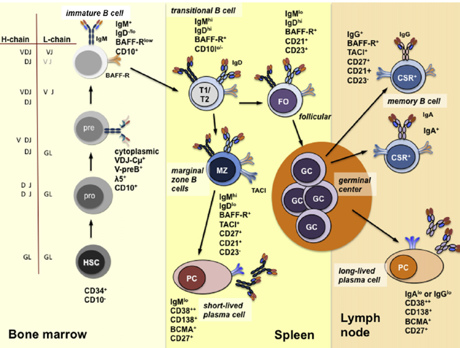 Figure 1: Development of B cells
B Cell Activation
Similar to T cells, B cell activation also requires two signals: The first signal is initiated when the BCR specifically binds to an antigen (T cell-dependent antigen, TD-Ag), transmitted by BCR-CD79a/b (BCR-Igα/β) and CD19/CD21/CD81. The second signal for B cell activation, also known as the co-stimulatory signal, is generated by the interaction of multiple pairs of co-stimulatory molecules between Th cells and B cells, the most important of which is CD40/CD40L. Additionally, cytokines secreted by Th cells can also promote B cell activation, proliferation, and differentiation. It is important to note that if only the first signal is present without the second, B cells will not only fail to activate but will also enter a state of anergic tolerance.
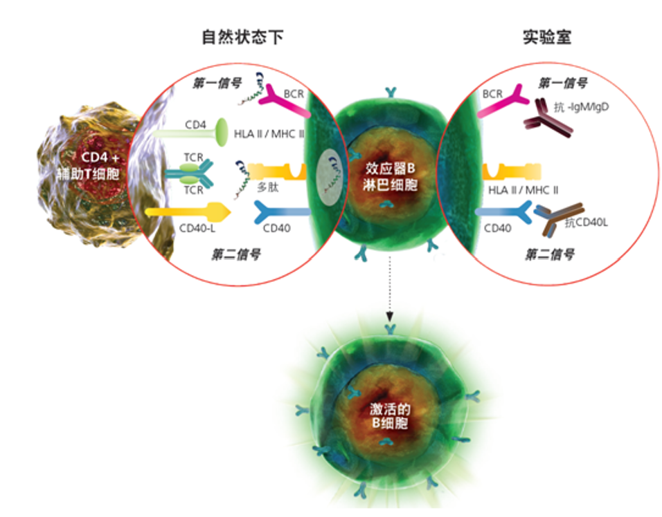
Figure 2: Activation of B cells
There is also another T cell-independent pathway for B cell activation, typically using T cell-independent antigens (TI-Ag), such as bacterial polysaccharides, polymeric proteins, and lipopolysaccharides. These antigens bind to molecules on the B cell surface rather than the cell receptor, aggregating these molecules. Consequently, the BCRs associated with these molecules also cluster together. This "polyclonal" B cell activation is independent of the cognate antigen recognized by the BCR; the BCR is merely drawn in. Based on the mechanism of B cell activation, TI antigens can be further divided into TI-1 antigens (LPS) and TI-2 antigens (bacterial cell wall and capsular polysaccharides). Both mature and immature B cells can be activated by TI-1 antigens, inducing the production of low-affinity IgM. TI-2 antigens can only activate mature B cells by extensively cross-linking mIg on B1 cells through their highly repetitive antigenic epitopes.
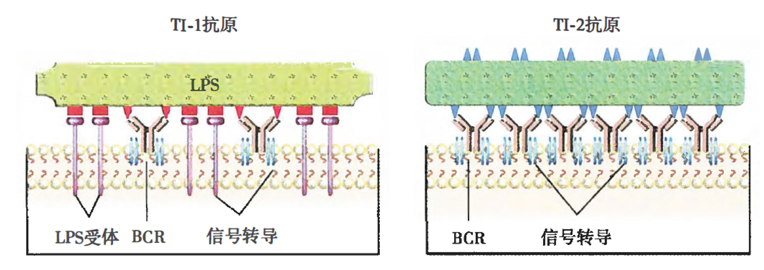
Figure 3: TI-1 and TI-2 antigens【2】: TI-1 antigens can bind to both BCR and mitogen receptors, activating B cells without T cell help; TI-2 antigens activate B cells by cross-linking multiple BCRs.
Example: In Vitro B Cell Activation and Expansion Protocol
(1) Preparation of Stromal Cells
Pre-seed a 10 cm cell culture dish with stromal cells expressing CD40L to serve as a feeder layer for B cell culture.
(2) B Cell Seeding
Seed B cells onto the feeder layer in the 10 cm dish (6 mL/dish) at a density of 100 cells/cm².
(3) Culture Conditions
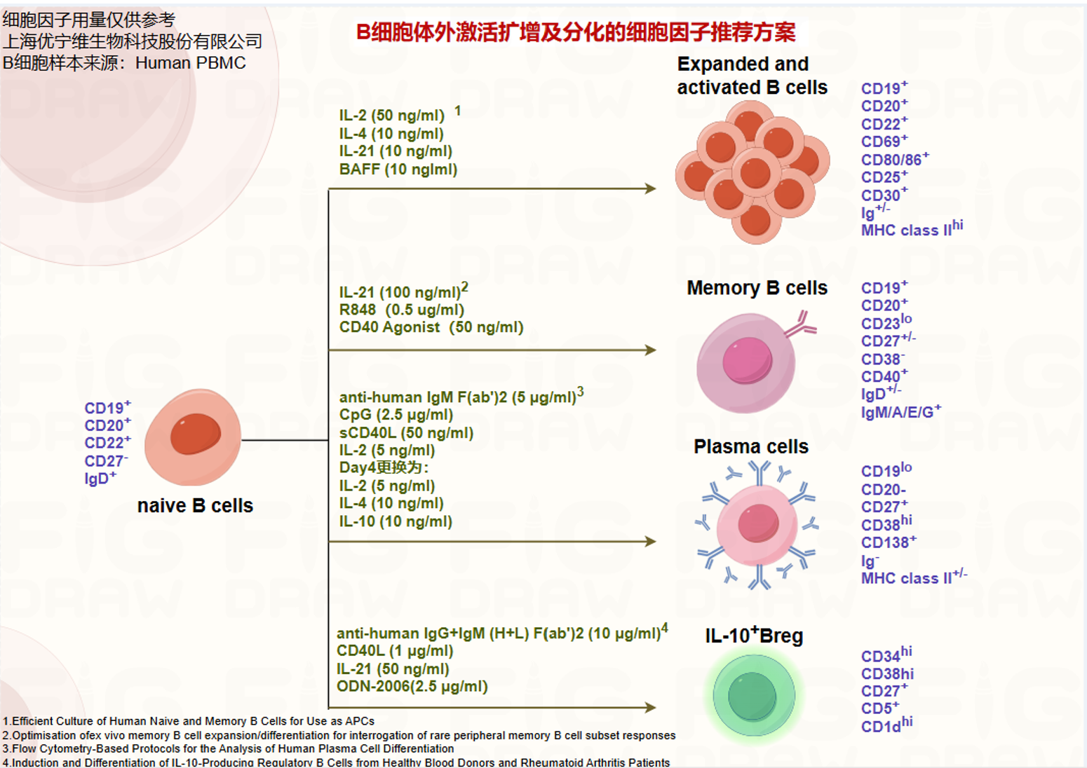
Use R5 medium (containing 5% human serum, 55 µM 2-mercaptoethanol (abs9592-100mL), 2 mM L-glutamine (abs9354-100mL), 100 U/mL penicillin, 100 µg/mL streptomycin (abs9244-100mL), 10 mM HEPES (abs42150994-100mL), 1 mM sodium pyruvate (abs9296-100mL), and 1% MEM non-essential amino acids (abs9298-100mL), supplemented with recombinant human IL-2 (50 ng/mL) (abs06485-25ug), IL-4 (10 ng/mL) (UA040026-50µg), IL-21 (10 ng/mL) (8879-IL-010/CF), and BAFF (10 ng/mL) (2149-BF-01M/CF). Culture B cells continuously for 8 days; the culture duration can be adjusted based on the experimental purpose. Formation of B cell clones can be observed during the culture period.
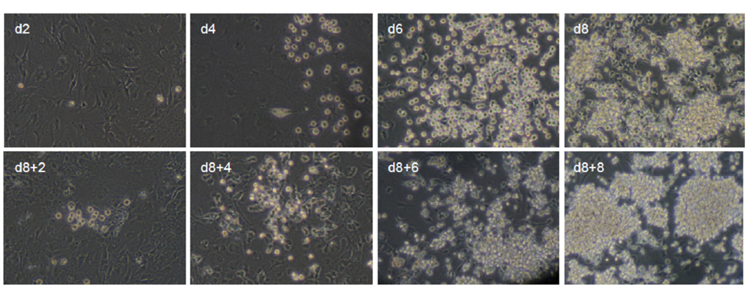 Figure 4: Clones formed by in vitro activation and expansion of human naïve B cells [3]
(4) Medium Change
On days 4 and 6, discard half of the old medium (avoid touching the cells at the bottom) and replace it with an equal volume of pre-warmed fresh R5 medium containing cytokines. When the culture exceeds 8 days, transfer the B cells to a new feeder layer on day 8. Similarly, replace half the medium on days 4 and 6 post-transfer. After the culture period, harvest the B cells for counting and subsequent experiments, or cryopreserve them in liquid nitrogen for later use.
B Cell Differentiation and Culture
Fully activated B cells, stimulated by both signals, acquire the capacity for proliferation and further differentiation. With the help of cytokines produced by Th cells, activated B cells proliferate to form germinal centers, where they undergo T-cell-dependent somatic hypermutation, Ig affinity maturation, and class switching, ultimately differentiating into plasma cells or memory B cells to exert humoral immune functions. Next, let's use the in vitro differentiation of memory B cells and plasma cells as examples.
01 In Vitro Differentiation and Proliferation of Memory B Cells【4】
a) Preparation of 3T3-msCD40L cells:
Seed 3T3-msCD40L cells in cell culture flasks and harvest them when the cells reach 80-90% confluency.
Seed 3T3-msCD40L cells in cell culture flasks and harvest them when the cells reach 80-90% confluency.
b) Cell Sorting:
Isolate CD19⁺IgM⁻IgA⁻IgD⁻ B cells using flow cytometric cell sorting.
Isolate CD19⁺IgM⁻IgA⁻IgD⁻ B cells using flow cytometric cell sorting.
c) Cell Culture:
Seed the sorted B cells into 384-well plates at a density of 1.3-4 cells per well. Each well contains 50 µL of complete medium. Add 100 µL of sterile water to the outermost wells of the 384-well plate to prevent evaporation.
Complete Medium Composition: 3.5 mL IL-2 (10000 U/mL) (UA040057-50µg) + 175 µL IL-21 (100 µg/mL) (abs05146-50ug) + 10 mL irradiated 3T3-msCD40L cells (total 3.5x10⁷ cells) + 336 mL Complete IMDM medium (abs9441-500mL).
Seed the sorted B cells into 384-well plates at a density of 1.3-4 cells per well. Each well contains 50 µL of complete medium. Add 100 µL of sterile water to the outermost wells of the 384-well plate to prevent evaporation.
Complete Medium Composition: 3.5 mL IL-2 (10000 U/mL) (UA040057-50µg) + 175 µL IL-21 (100 µg/mL) (abs05146-50ug) + 10 mL irradiated 3T3-msCD40L cells (total 3.5x10⁷ cells) + 336 mL Complete IMDM medium (abs9441-500mL).
d) Culture Conditions:
Culture cells at 37°C and 5% CO₂ for 13-14 days.
Culture cells at 37°C and 5% CO₂ for 13-14 days.
e) Monitoring and Supernatant Collection:
Monitor under a microscope periodically. Around day 10, expanding B cell clones can be observed. Measure the IgG concentration in the supernatant on day 12 (abs551021-96T, Human IgG ELISA Kit) to analyze the differentiation efficiency of memory B cells.
Monitor under a microscope periodically. Around day 10, expanding B cell clones can be observed. Measure the IgG concentration in the supernatant on day 12 (abs551021-96T, Human IgG ELISA Kit) to analyze the differentiation efficiency of memory B cells.
02 In Vitro Differentiation and Proliferation of Plasma Cells【5】
a) Cell Preparation:
Isolate peripheral blood mononuclear cells (PBMCs) from healthy donor blood using density gradient centrifugation (17144003-1, or abs930-200mL).
Isolate peripheral blood mononuclear cells (PBMCs) from healthy donor blood using density gradient centrifugation (17144003-1, or abs930-200mL).
b) B Cell Sorting:
Isolate total B cells from PBMCs using magnetic bead-based cell separation (130-101-638).
Isolate total B cells from PBMCs using magnetic bead-based cell separation (130-101-638).
c) In Vitro B Cell Culture:
Seed B cells in 48-well plates at a density of 2.5×10⁵ cells/mL. B cells can be labeled with CellTrace Violet (CTV) (48444) to track cell divisions.
Seed B cells in 48-well plates at a density of 2.5×10⁵ cells/mL. B cells can be labeled with CellTrace Violet (CTV) (48444) to track cell divisions.
d) Stimulation Conditions:
Use 4 different stimulation conditions, including CpG ODN 2395 (abs9983-5mg), soluble CD40 ligand (sCD40L), recombinant human IL-2 and IL-10 (abs06283-50ug), etc., to induce B cell differentiation into plasma cells.
Use 4 different stimulation conditions, including CpG ODN 2395 (abs9983-5mg), soluble CD40 ligand (sCD40L), recombinant human IL-2 and IL-10 (abs06283-50ug), etc., to induce B cell differentiation into plasma cells.
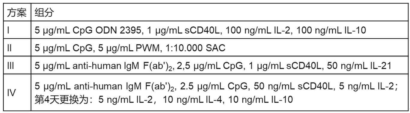
Figure 5: Exploring experimental protocols for in vitro plasma cell differentiation
e) Flow Cytometry Analysis:
On days 3.5 and 6, analyze B cell surface markers such as CD38 (130-113-431), CD20 (556633), and transcription factor expression by flow cytometry.
On days 3.5 and 6, analyze B cell surface markers such as CD38 (130-113-431), CD20 (556633), and transcription factor expression by flow cytometry.
f) ELISA Detection:
On day 6 of culture, collect the supernatant for ELISA to detect IgG production.
On day 6 of culture, collect the supernatant for ELISA to detect IgG production.
g) Optimal Plasma Cell Differentiation Protocol:
Determine Protocol IV as the best among the four based on flow cytometry and ELISA results.
Determine Protocol IV as the best among the four based on flow cytometry and ELISA results.
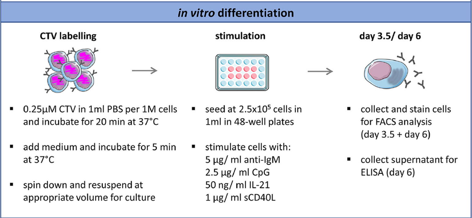
Figure 6: In vitro plasma cell differentiation experimental protocols and detection
03 Memory B Cell Differentiation into Plasma Cells【6】
a) B Cell Sorting:
Isolate CD27⁺ memory B cells from healthy donor PBMCs using magnetic bead-based cell separation (130-051-601).
Isolate CD27⁺ memory B cells from healthy donor PBMCs using magnetic bead-based cell separation (130-051-601).
b) Medium Preparation:
Use RPMI-1640 medium (abs9484-500mL) supplemented with L-glutamine, 1% penicillin/streptomycin, and 10% fetal bovine serum (abs972-500mL or SV30208.02).
Use RPMI-1640 medium (abs9484-500mL) supplemented with L-glutamine, 1% penicillin/streptomycin, and 10% fetal bovine serum (abs972-500mL or SV30208.02).
c) Cell Culture and Stimulation:
Add feeder cells (irradiated CD154⁺ HEK-293T) at a ratio of 1:1 to 1:50. Then, seed the sorted memory B cells into 96-well plates at a density of 8×10³ cells/mL for Ig quantification in supernatant, or into 24-well plates at a density of 1×10⁶ cells/mL for phenotypic analysis by flow cytometry.
Use different combinations of stimulants to induce memory B cell differentiation into plasma cells, including IL-2 (100-1000 U/mL), IL-6 (10-100 ng/mL) (UA040052-50µg), IL-15 (10-100 ng/mL) (abs05147-10ug), IL-21 (10-100 ng/mL), APRIL (10-100 ng/mL), BAFF (10-100 ng/mL), CpG ODN2006 (0.25-10 µg/mL) (130-100-105), R848 (0.25-5 µg/mL), and pokeweed mitogen (5-100 ng/mL) (abs810694-50mg). Culture cells at 37°C and 5% CO₂ for 5-10 days.
Add feeder cells (irradiated CD154⁺ HEK-293T) at a ratio of 1:1 to 1:50. Then, seed the sorted memory B cells into 96-well plates at a density of 8×10³ cells/mL for Ig quantification in supernatant, or into 24-well plates at a density of 1×10⁶ cells/mL for phenotypic analysis by flow cytometry.
Use different combinations of stimulants to induce memory B cell differentiation into plasma cells, including IL-2 (100-1000 U/mL), IL-6 (10-100 ng/mL) (UA040052-50µg), IL-15 (10-100 ng/mL) (abs05147-10ug), IL-21 (10-100 ng/mL), APRIL (10-100 ng/mL), BAFF (10-100 ng/mL), CpG ODN2006 (0.25-10 µg/mL) (130-100-105), R848 (0.25-5 µg/mL), and pokeweed mitogen (5-100 ng/mL) (abs810694-50mg). Culture cells at 37°C and 5% CO₂ for 5-10 days.
d) Optimal Expansion Conditions:
IL-21 concentration: 100 ng/mL. R848 concentration: 0.5 µg/mL. CD40 stimulation: Use CD154⁺ HEK293T cells as feeder cells at a 1:1 ratio with memory B cells.
IL-21 concentration: 100 ng/mL. R848 concentration: 0.5 µg/mL. CD40 stimulation: Use CD154⁺ HEK293T cells as feeder cells at a 1:1 ratio with memory B cells.
Here is a summary of common B cell differentiation protocols compiled for reference.
Related Products Mentioned in this Section
Promotes B cell survival and proliferation
Promotes B cell class switching to IgE and IgG1
Acts synergistically with IL-4 to promote class switching
Promotes B cell survival and differentiation
Promotes B cell survival, maturation, and class switching
A TLR7/8 agonist that promotes B cell activation, proliferation, and differentiation
Binds to CD40, mimicking the interaction between T cells and B cells
B Cell Identification
B cell identification is typically performed by flow cytometry. The different B cell subtypes and their markers are illustrated in Figure 8【7】.
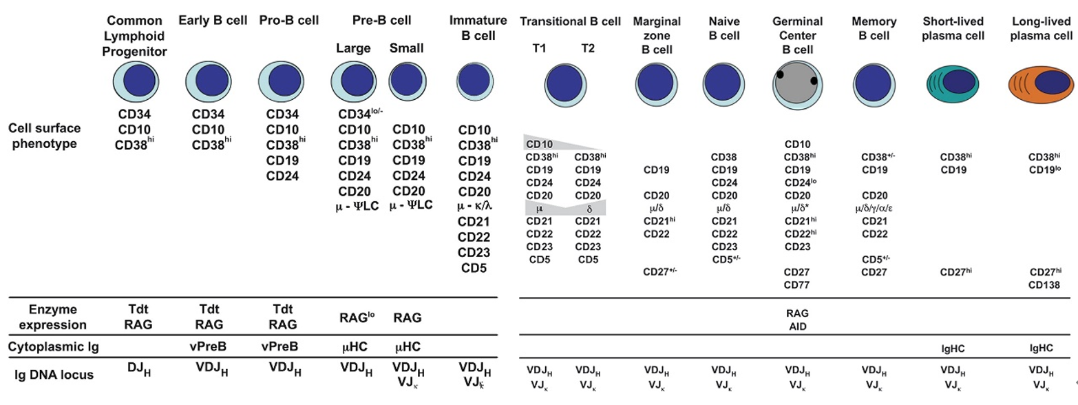
Figure 8: Human B cell surface markers [7]
Related Products Mentioned in this Section
Applications: FC, MICS, IF, IHC
Application: FC
Hot Topics in B Cell Research
There are many hot research topics related to B cells, such as BCR function, Breg cells, and B cells in tertiary lymphoid structures. Taking B cells and Tertiary Lymphoid Structures (TLS) as an example: TLS are ectopic lymphoid organs that form in inflamed tissues under chronic antigenic stimulation. Mature TLS contain T cell zones and B cell zones, with T cells located peripherally and B cells centrally. B cells within TLS can acquire antigen from follicular dendritic cells (FDCs) and produce antibodies against the relevant antigens. Thus, TLS can drive specific immune responses within tumors. Research can focus on B cell types within TLS, chemokines secreted by B cells (LXLBH10-1, Human 10-Plex Detection Panel), antibody isotypes, etc. For instance, using transcriptomics combined with mIHC (abs50013-20T, 5-Color Multiplex Fluorescent Immunohistochemistry Staining Kit) to investigate IgG class switching in B cells within TLS: IgG isotypes (IGHG1-4) are enriched in TLS+ tumors; IgA isotypes (IGHA1-2) are enriched in TLS- tumors【8】.
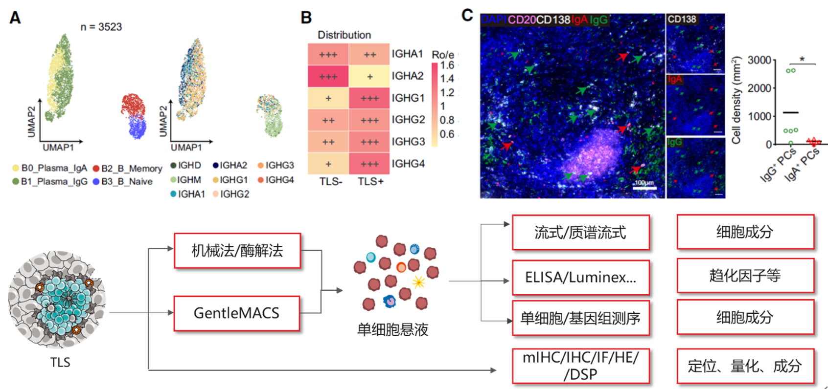 Figure 9: Summary of hot research directions in B cell biology
Related Products Mentioned in this Section
Up to 9 markers (10 colors including DAPI) multiplex fluorescent IHC service, offering whole-slide scanning and quantitative analysis
References:
1. Pieper, K., B. Grimbacher, and H. Eibel, B-cell biology and development. Journal of Allergy and Clinical Immunology, 2013. 131(4): p. 959-971.
2. 医学免疫学_第9版
3. Su, K.-Y., et al., Efficient Culture of Human Naive and Memory B Cells for Use as APCs. The Journal of Immunology, 2016. 197(10): p. 4163-4176.
4. Huang, J., et al., Isolation of human monoclonal antibodies from peripheral blood B cells. Nature Protocols, 2013. 8(10): p. 1907-1915.
5. Khoenkhoen, S., et al., Flow Cytometry-Based Protocols for the Analysis of Human Plasma Cell Differentiation. Frontiers in Immunology, 2020. 11.
6. Muir, L., et al., Optimisation of ex vivo memory B cell expansion/differentiation for interrogation of rare peripheral memory B cell subset responses. Wellcome Open Research, 2017. 2.
7. Levesque, M.C. and E.W. St. Clair, B cell--directed therapies for autoimmune disease and correlates of disease response and relapse. Journal of Allergy and Clinical Immunology, 2008. 121(1): p. 13-21.
8. Zhang, Y., et al., CCL19-producing fibroblasts promote tertiary lymphoid structure formation enhancing anti-tumor IgG response in colorectal cancer liver metastasis. Cancer Cell, 2024. 42(8): p. 1370-1385.e9.
2. 医学免疫学_第9版
3. Su, K.-Y., et al., Efficient Culture of Human Naive and Memory B Cells for Use as APCs. The Journal of Immunology, 2016. 197(10): p. 4163-4176.
4. Huang, J., et al., Isolation of human monoclonal antibodies from peripheral blood B cells. Nature Protocols, 2013. 8(10): p. 1907-1915.
5. Khoenkhoen, S., et al., Flow Cytometry-Based Protocols for the Analysis of Human Plasma Cell Differentiation. Frontiers in Immunology, 2020. 11.
6. Muir, L., et al., Optimisation of ex vivo memory B cell expansion/differentiation for interrogation of rare peripheral memory B cell subset responses. Wellcome Open Research, 2017. 2.
7. Levesque, M.C. and E.W. St. Clair, B cell--directed therapies for autoimmune disease and correlates of disease response and relapse. Journal of Allergy and Clinical Immunology, 2008. 121(1): p. 13-21.
8. Zhang, Y., et al., CCL19-producing fibroblasts promote tertiary lymphoid structure formation enhancing anti-tumor IgG response in colorectal cancer liver metastasis. Cancer Cell, 2024. 42(8): p. 1370-1385.e9.
Disclaimer: This article partially utilizes artificial intelligence assistance in its creation. If any content involves copyright or intellectual property issues, please let us know and we promise to verify and remove it as soon as possible.
Purchase recombinant protein, choose Nanjing UA-Bio
UA protein focuses on providing various protein reagents, raw materials, and services required for drug research and development, cell therapy, gene therapy, and basic scientific research, including drug target proteins, immune checkpoint proteins, cytokines, tool enzymes, customized protein expression, and full-length transmembrane protein development. Youai is committed to providing customers with high-quality products and professional services, and building a high-tech enterprise with international competitiveness.
Target proteins | membrane proteins | cytokines | enzymes | viral antigens | protein customization
Nanjing UA Biotechnology Co., Ltd. Email:order@ua-bio.com Phone:0571-87565022


Link:





©2021-2026 all rights reserved UA Biotechnology Co.,Ltd.

网站备案号 浙ICP备2022019033号-2 浙公网安备33010202004867号
 Business license (three certificates in one)
 Tel:+86-0571-87565022
E-mail:order@ua-bio.com
All products sold on this website shall not be used for clinical diagnosis or treatment of humans or animals, and shall only be used for non-medical purposes such as industrial or scientific research.
