

NK Cell Culture Guide
Views:5 


Natural killer cells (NK cells) are the third major type of lymphocytes, apart from T cells and B cells. They can spontaneously recognize abnormal cells in the body, rapidly eliminate them through cytotoxic effects, and produce various pro-inflammatory factors and chemokines to recruit and activate other immune cells, thereby initiating adaptive immune responses.
- Recent Advances
Recent Advances
NK Cell Culture Guide
Natural killer (NK) cells are the third major type of lymphocytes besides T cells and B cells. They can spontaneously recognize abnormal cells in the body and rapidly eliminate them through cytotoxic effects, while producing various pro-inflammatory factors and chemokines to recruit and activate other immune cells to initiate adaptive immune responses.
NK Cell Sources
NK cells originate from bone marrow lymphoid stem cells, and their differentiation and development depend on the bone marrow microenvironment. They are mainly distributed in the bone marrow, peripheral blood, liver, spleen, lungs, and lymph nodes, accounting for about 5-20% of peripheral lymphocytes. In the blood, bone marrow, spleen, and lungs, NK cells are primarily mature (CD56dimCD16+) and terminally differentiated (CD56dimCD16+CD57+) subsets, with higher effector functions. In lymph nodes, tonsils, and the intestines, NK cells are less frequent and consist mainly of immature (CD56brightCD16−) subsets, which have lower effector capabilities[1].
Therefore, the sources of NK cells for in vitro culture can be solid tissues (bone marrow, spleen, liver, lymph nodes, etc.), blood, commercially available primary NK cells (2W-502), or NK cell lines (such as NK-92).

Figure 1 NK Cell Development and Distribution[2]
Related Products in the Article
Mouse spleen tissue dissociation kit for efficient preparation of single-cell suspensions
Used for primary cell preparation from tissues such as heart, bone, liver, thyroid, salivary glands, and cartilage
For digesting islet cells and experimental samples requiring high integrity of cell surface receptors
Rapid and gentle negative selection of NK cells from human PBMCs
In Vitro Expansion and Culture of NK Cells
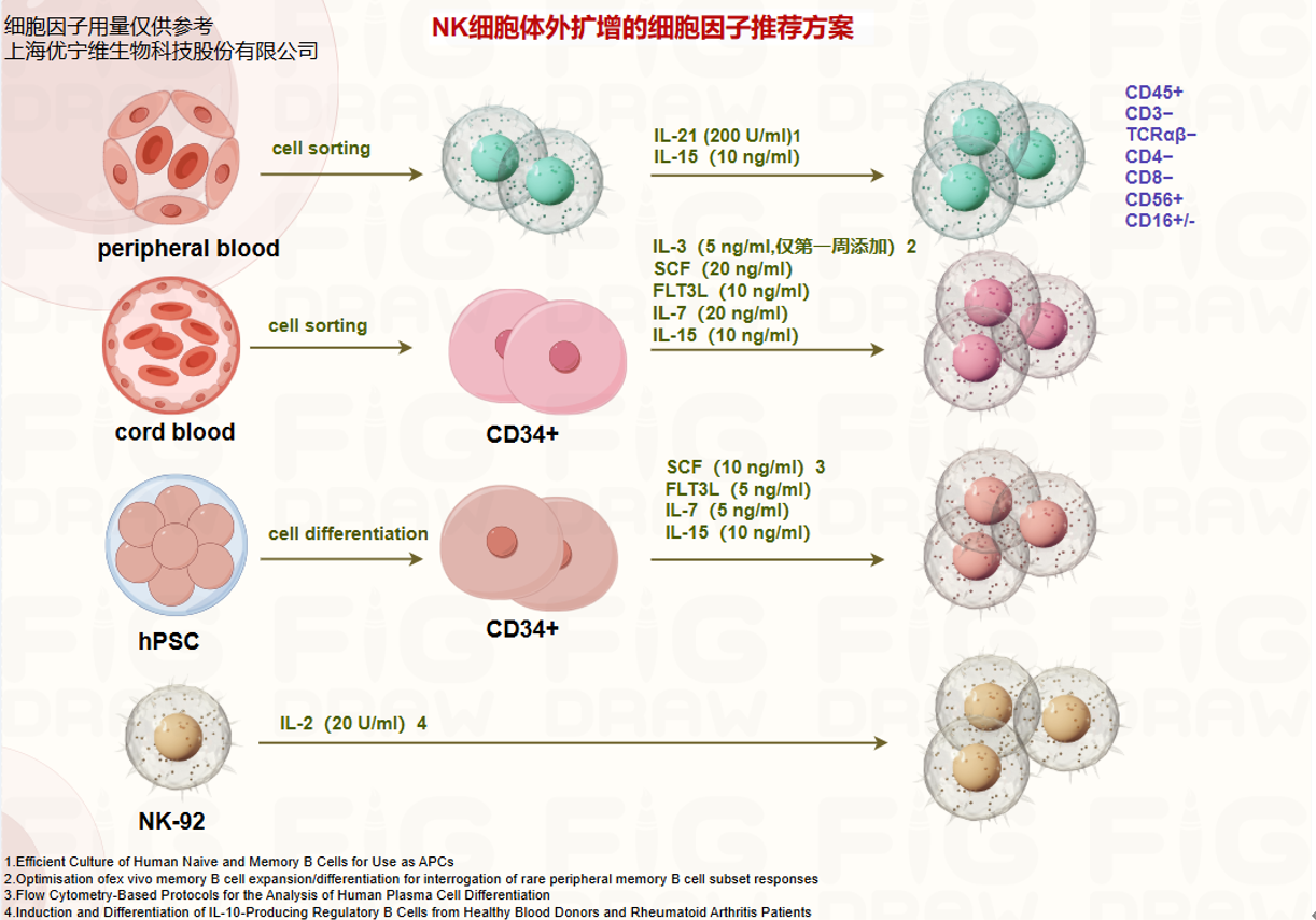
NK cells also face many challenges in vitro: NK cells account for a low proportion in peripheral blood, and PBMC-based expansion yields low NK cell ratios; NK cells are difficult to expand and prone to exhaustion, requiring serum or feeder cells for culture; expanded NK cells often struggle to maintain their cytotoxic functions. Therefore, different NK cell culture factor combinations can be used to address these challenges: IL-2, IL-12, and IL-15 promote NK cell differentiation and expansion; IL-12 and IL-18 enhance NK cell antigen sensitivity; IL-21 enhances NK cell cytotoxicity.
Next, we will detail four case studies:
1. Peripheral Blood-Derived NK Cells (PB-Derived NK Cells)
① Adjust the density of NK cells isolated from human whole blood (130-127-695) to 1×10⁶ cells/mL and resuspend in X-VIVO 15 medium (04-418Q) containing 5% serum (SV30208.02);
② Add 200 U/mL IL-2 (UA040057-50μg), 10 ng/mL IL-15 (UA040010-50μg), and 1×10⁶ cells/mL of K562 cells (inactivated by mitomycin treatment). During the 15-day expansion, NK cells are passaged every 2-3 days, with cytokines and freshly prepared K562 cells replenished[3].
2. Umbilical Cord Blood-Derived NK Cells (UCB-Derived NK Cells)
① Isolate CD34+ cells: Use CD34-positive selection magnetic beads (130-046-702) to separate CD34-expressing hematopoietic stem/progenitor cells from umbilical cord blood;
② Resuspend the isolated CD34+ cells in hematopoietic stem cell differentiation medium (DMEM/F12 (SH30271.01) + 20% heat-inactivated human AB serum + 5 ng/mL sodium selenite, 50 µM ethanolamine, 20 mg/mL ascorbic acid + 2 mM L-glutamine (abs9354-100mL), 1% penicillin-streptomycin (SV30010), 25 µM β-mercaptoethanol (abs9592-100mL)) and seed them onto irradiated OP9-DLL4 cells, supplemented with the following cytokines: 5 ng/mL IL-3 (only in the first week), 20 ng/mL SCF (UA040115-1mg), 10 ng/mL IL-15 (UA040010-1mg), 10 ng/mL FLT3L (UA040011-1mg), 20 ng/mL IL-7 (UA040118-1mg);
③ Replace the medium weekly to provide continuous nutrient and growth factor support. After about 28 to 35 days, flow cytometry can detect changes in cell surface markers. When cells develop into a CD45+CD56+CD33−CD3− phenotype, they have differentiated into NK cells[4].
3. hPSC-Derived NK Cells[5]
1) Hematopoietic Progenitor Cell Differentiation
① Use OP9 bone marrow stromal cells as supporting cells and co-culture with H1 cells (α-MEM + 20% FBS) to induce hematopoietic progenitor cell differentiation;
② After about 12 days of co-culture, many differentiated colonies can be observed, and a small portion of CD34+ cells can be detected.
① Use OP9 bone marrow stromal cells as supporting cells and co-culture with H1 cells (α-MEM + 20% FBS) to induce hematopoietic progenitor cell differentiation;
② After about 12 days of co-culture, many differentiated colonies can be observed, and a small portion of CD34+ cells can be detected.
2) Induction of NK Cell Differentiation
① Harvest cells obtained after the "hematopoietic progenitor cell differentiation" phase and co-culture them with OP9 cells expressing the Notch ligand Delta-like-1 (OP9-DLL1 cells);
② In α-MEM medium (abs9506-500mL) containing 20% FBS (abs972-500mL), 10 ng/mL SCF (abs05620-50ug), 5 ng/mL FLT3L (abs05148-50ug), 5 ng/mL IL-7 (abs04991-100ug), and 10 ng/mL IL-15 (abs00818-50ug), perform such co-culture weekly.
③ After the first cycle of co-culture (day 19), most cells remain adherent.
④ During the second cycle (day 26), some bright, round, semi-suspended cells begin to appear.
⑤ During the third cycle (day 33), these cells gradually detach and aggregate in the center of the culture well.
⑥ During the fourth cycle (day 40), more suspended cells appear, exhibiting small, round, bright lymphocyte morphology; flow cytometry can identify these cells as CD45+CD56+CD3−TCRαβ−CD4−CD8−, i.e., NK cells.
① Harvest cells obtained after the "hematopoietic progenitor cell differentiation" phase and co-culture them with OP9 cells expressing the Notch ligand Delta-like-1 (OP9-DLL1 cells);
② In α-MEM medium (abs9506-500mL) containing 20% FBS (abs972-500mL), 10 ng/mL SCF (abs05620-50ug), 5 ng/mL FLT3L (abs05148-50ug), 5 ng/mL IL-7 (abs04991-100ug), and 10 ng/mL IL-15 (abs00818-50ug), perform such co-culture weekly.
③ After the first cycle of co-culture (day 19), most cells remain adherent.
④ During the second cycle (day 26), some bright, round, semi-suspended cells begin to appear.
⑤ During the third cycle (day 33), these cells gradually detach and aggregate in the center of the culture well.
⑥ During the fourth cycle (day 40), more suspended cells appear, exhibiting small, round, bright lymphocyte morphology; flow cytometry can identify these cells as CD45+CD56+CD3−TCRαβ−CD4−CD8−, i.e., NK cells.
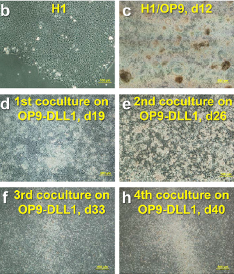
Morphological Monitoring During hPSC-Induced NK Cell Differentiation[5]
4. NK Cell Line: NK-92
The culture of the NK-92 cell line is much simpler compared to primary NK cells. Adjust the NK cell density to 2.5×10⁵/mL and resuspend in complete medium (RPMI 1640 (SH30809.01) + 5% FBS (SH30406.05) + 2 mM L-glutamine (abs9354-100mL) + 10 U/mL penicillin-streptomycin (SV30010) + 20 U/mL IL-2 (UA040057-50μg))[6], replacing the complete medium every 2-3 days.
We have also summarized the in vitro expansion and culture protocols for NK cells from various sources for reference~
NK Cell Identification
The most common method for identifying NK cell subtypes is flow cytometry. For basic information on flow cytometry identification, please refer to "Immunocyte Culture Techniques: Phenotyping and Identification." Common human NK cell identification markers are: CD3−CD56+CD16+/−. Human NK cells are often divided into two subsets: CD56++CD16+/− and CD56dimCD16++. For peripheral blood or other non-purified NK cell samples, CD3 must also be detected to exclude a special type of T cell—NKT (CD3+CD56+). Common mouse NK cell identification markers are CD3e−CD49b+NK1.1+NKp46+, but the choice of NK1.1 and CD49b depends on the mouse strain: NK1.1 is expressed in C57BL, FVB/N, NZB but not in AKR, BALB/c, CBA/J, C3H, C57BR, C58, DBA/1, DBA/2, NOD, SJL, 129. CD49b is also expressed in thymic epithelial cells, B cells, monocytes, etc. For NK cell identification, antibodies with the antigen clone DX5 are recommended for stronger specificity, as they are expressed in almost all mouse strains except NOD.
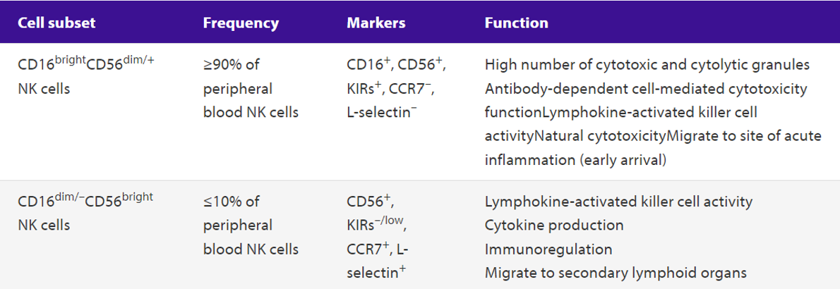
Human NK Cell Subtypes, Proportions, Markers, Distribution, and Function (Source: Miltenyi Biotec)
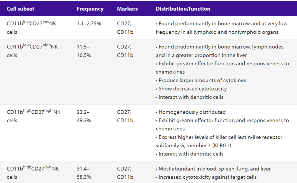
Mouse NK Cell Subtypes, Proportions, Markers, Distribution, and Function (Source: Miltenyi Biotec)
Related Products in the Article
Hot Research Topics in NK Cells
There are many hot research topics in NK cells, such as NK cell subtypes and functions in the tumor microenvironment, NK cell exhaustion, tumor immune escape, interactions between NK cells and other cells, CAR-NK, and NK cell functional studies. For example, a 2024 study co-cultured NK cells, T cells, and tumor cells, added a trispecific antibody, and used confocal microscopy imaging and ELISA to detect cytokine levels in the co-culture supernatant to observe whether the trispecific antibody could enhance the interaction between NK cells, T cells, and tumor cells and stimulate the release of cytotoxic granules (Figure 1)[7].

Co-culture of NK Cells, T Cells, and Tumor Cells[7]
Related Products in the Article
References:
1. Dogra, P., et al., Tissue Determinants of Human NK Cell Development, Function, and Residence. Cell, 2020. 180(4): p. 749-763.e13.
2. Crinier, A., et al., SnapShot: Natural Killer Cells. Cell, 2020. 180(6): p. 1280-1280.e1.
3. Li, F., et al., CCL5-armed oncolytic virus augments CCR5-engineered NK cell infiltration and antitumor efficiency. Journal for ImmunoTherapy of Cancer, 2020. 8(1).
4. Goldenson, B.H., et al., Umbilical Cord Blood and iPSC-Derived Natural Killer Cells Demonstrate Key Differences in Cytotoxic Activity and KIR Profiles. Frontiers in Immunology, 2020. 11.
5. Zeng, J., et al., Generation of "Off-the-Shelf" Natural Killer Cells from Peripheral Blood Cell-Derived Induced Pluripotent Stem Cells. Stem Cell Reports, 2017. 9(6): p. 1796-1812.
6. Törnroos, H., H. Hägerstrand, and C. Lindqvist, Culturing the Human Natural Killer Cell Line NK-92 in Interleukin-2 and Interleukin-15 -- Implications for Clinical Trials. Anticancer Research, 2018. 39(1): p. 107-112.
7. Ye, Q.N., et al., Orchestrating NK and T cells via tri-specific nano-antibodies for synergistic antitumor immunity. Nat Commun, 2024. 15(1): p. 6211.
1. Dogra, P., et al., Tissue Determinants of Human NK Cell Development, Function, and Residence. Cell, 2020. 180(4): p. 749-763.e13.
2. Crinier, A., et al., SnapShot: Natural Killer Cells. Cell, 2020. 180(6): p. 1280-1280.e1.
3. Li, F., et al., CCL5-armed oncolytic virus augments CCR5-engineered NK cell infiltration and antitumor efficiency. Journal for ImmunoTherapy of Cancer, 2020. 8(1).
4. Goldenson, B.H., et al., Umbilical Cord Blood and iPSC-Derived Natural Killer Cells Demonstrate Key Differences in Cytotoxic Activity and KIR Profiles. Frontiers in Immunology, 2020. 11.
5. Zeng, J., et al., Generation of "Off-the-Shelf" Natural Killer Cells from Peripheral Blood Cell-Derived Induced Pluripotent Stem Cells. Stem Cell Reports, 2017. 9(6): p. 1796-1812.
6. Törnroos, H., H. Hägerstrand, and C. Lindqvist, Culturing the Human Natural Killer Cell Line NK-92 in Interleukin-2 and Interleukin-15 -- Implications for Clinical Trials. Anticancer Research, 2018. 39(1): p. 107-112.
7. Ye, Q.N., et al., Orchestrating NK and T cells via tri-specific nano-antibodies for synergistic antitumor immunity. Nat Commun, 2024. 15(1): p. 6211.
```
Disclaimer: This article partially utilizes artificial intelligence assistance in its creation. If any content involves copyright or intellectual property issues, please let us know and we promise to verify and remove it as soon as possible.
Purchase recombinant protein, choose Nanjing UA-Bio
UA protein focuses on providing various protein reagents, raw materials, and services required for drug research and development, cell therapy, gene therapy, and basic scientific research, including drug target proteins, immune checkpoint proteins, cytokines, tool enzymes, customized protein expression, and full-length transmembrane protein development. Youai is committed to providing customers with high-quality products and professional services, and building a high-tech enterprise with international competitiveness.
Target proteins | membrane proteins | cytokines | enzymes | viral antigens | protein customization
Nanjing UA Biotechnology Co., Ltd. Email:order@ua-bio.com Phone:0571-87565022


Link:





©2021-2026 all rights reserved UA Biotechnology Co.,Ltd.

网站备案号 浙ICP备2022019033号-2 浙公网安备33010202004867号
 Business license (three certificates in one)
 Tel:+86-0571-87565022
E-mail:order@ua-bio.com
All products sold on this website shall not be used for clinical diagnosis or treatment of humans or animals, and shall only be used for non-medical purposes such as industrial or scientific research.
