

Neuron and Glial Cell Culture Guide
Views:7 


Neurons and neuroglial cells are the basic units that constitute the structure and function of the nervous system.
- Recent Advances
Recent Advances
Neuron and Glial Cell Culture Guide
Neurons and glial cells are the fundamental units of the nervous system's structure and function. Neurons are responsible for receiving, processing, transmitting, and storing information. Glial cells provide structural support to neurons and participate in immune responses. For example, astrocytes maintain the blood-brain barrier and aid in injury repair; microglia clear pathogens and participate in inflammatory responses; oligodendrocytes form myelin sheaths around axons to accelerate nerve signal conduction. Research on neurons and glial cells helps explore the pathogenesis and treatment of nervous system diseases.
In this issue, we focus on the foundation of neuroscience research—the guide to neural cell culture. Next, Xiao You will take you through six essential skills for studying different neural cells: neural cell development, primary cell extraction, neural cell sorting, neural cell culture, neural cell identification, and popular neural-related research. The article is packed with valuable information and takes about 15 minutes to read. We recommend bookmarking it!
1. Neural Cell Development
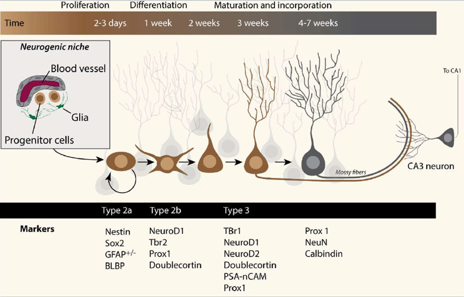
Neural stem cells proliferate in a microenvironment composed of blood vessels and astrocytes, producing neural progenitor cells. These progenitor cells develop into neuroblasts and begin migrating, gradually differentiating into immature neurons. After 2-3 weeks, their morphology and function further develop. Immature neurons continue to mature, becoming fully functional neurons after 4 weeks, some of which can establish connections with the CA3 region of the hippocampus to perform neural functions[1].
2. Primary Cell Extraction
Primary neurons are mainly extracted from the central nervous system (CNS) tissues of embryonic or newborn animals. The specific extraction site depends on the type of neuron being studied. For example, glutamatergic excitatory neurons can be extracted from the cerebral cortex of rat/mouse embryos (E15-E18); CA1/CA3 pyramidal neurons can be extracted from the hippocampus of rat/mouse embryos (E15-E18). The developmental stage of the tissue must be considered. For instance, during the embryonic period (E15-E19), glial cells have not proliferated extensively, so neuron purity exceeds 90%. In contrast, during the neonatal period (P0-P7), enzymatic digestion combined with differential adhesion is required to remove glial cells. Neurons are highly susceptible to contamination and must be handled in a sterile environment with antibiotics. Trypsin treatment should not exceed 15-20 minutes (37°C), and the entire process from extraction to inoculation should be completed within 2-3 hours to avoid cell death. Before inoculation, the culture surface must be coated with substrates such as poly-L-lysine (PLL) or laminin.
Astrocytes are typically extracted from the cerebral cortex, hippocampus, or spinal cord of newborn rats/mice (P0-P4), as this period has fewer neurons and oligodendrocyte precursor cells, making it easier to obtain high-purity astrocytes. Microglia are usually extracted from the cerebral cortex, midbrain, or brainstem of newborn to juvenile rats/mice (P0-P14), as this is the peak period for microglial settlement and proliferation. Adult brains can also be used, but cell yield, viability, and purity may decrease. Oligodendrocytes are generally extracted from the cerebral cortex, corpus callosum, or spinal cord of newborn rats/mice (P0-P3), as oligodendrocyte precursor cells (OPCs) are abundant in white and gray matter around birth.
Steps for Primary Neural Cell Extraction (using newborn mouse brain as an example):
1. Prepare enzyme solutions: Precisely prepare Enzyme Mix 1 and Enzyme Mix 2 according to the instructions of the dissociation kit;
2. Tissue extraction and weighing: Collect mouse brain tissue and weigh it in 1 mL of calcium- and magnesium-free HBSS (SH30588.01);
3. Transfer up to 400 mg of tissue to a gentleMACS C Tube (130-093-237) pre-filled with the required volume of Enzyme Mix 1;
4. Add 30 µL of Enzyme Mix 2 to the C Tube (130-093-237) and mix gently by inversion;
5. Tighten the cap, invert the tube, and attach it to the gentleMACS Octo tissue dissociator (130-096-427), ensuring the sample is in the rotor/stator area. Run the preset program 37C_NTDK_1;
6. After the program ends, briefly centrifuge to collect the sample at the bottom of the tube;
7. Filter the cell suspension through a pre-wetted 70 µm MACS SmartStrainer (130-098-462) into a 50 mL centrifuge tube;
8. Rinse the filter and cells with 10 mL of calcium- and magnesium-containing HBSS (SH30030.02);
9. Discard the filter and centrifuge the cell suspension at 300×g for 10 minutes. Carefully aspirate and discard the supernatant;
10. Resuspend the cell pellet in an appropriate buffer such as PBS (SH30258.01) for subsequent experiments.
2. Tissue extraction and weighing: Collect mouse brain tissue and weigh it in 1 mL of calcium- and magnesium-free HBSS (SH30588.01);
3. Transfer up to 400 mg of tissue to a gentleMACS C Tube (130-093-237) pre-filled with the required volume of Enzyme Mix 1;
4. Add 30 µL of Enzyme Mix 2 to the C Tube (130-093-237) and mix gently by inversion;
5. Tighten the cap, invert the tube, and attach it to the gentleMACS Octo tissue dissociator (130-096-427), ensuring the sample is in the rotor/stator area. Run the preset program 37C_NTDK_1;
6. After the program ends, briefly centrifuge to collect the sample at the bottom of the tube;
7. Filter the cell suspension through a pre-wetted 70 µm MACS SmartStrainer (130-098-462) into a 50 mL centrifuge tube;
8. Rinse the filter and cells with 10 mL of calcium- and magnesium-containing HBSS (SH30030.02);
9. Discard the filter and centrifuge the cell suspension at 300×g for 10 minutes. Carefully aspirate and discard the supernatant;
10. Resuspend the cell pellet in an appropriate buffer such as PBS (SH30258.01) for subsequent experiments.
Some related products mentioned in the text:
【Tissue Preservation】
MACS® Freezing Solution, 130-129-552
Tissue preservation solution for storing brain tissue before dissociation
【Tissue Dissociator】
gentleMACS 25 C Tubes, 130-093-237
gentleMACS™ Octo Dissociator with Heaters, 130-096-427
【Tissue Dissociation Kits】
Adult Brain Dissociation Kit, mouse and rat, 130-107-677
Dissociation kit for adult mouse/rat (P > 7) brain tissue
Neural Tissue Dissociation Kit (P), 130-092-628
Dissociation kit for newborn mouse/rat (P ≤ 7) brain tissue, papain-based
Neural Tissue Dissociation Kit (T), 130-093-231
Dissociation kit for newborn mouse/rat (P ≤ 7) brain tissue, trypsin-based
Neural Tissue Dissociation Kit---Postnatal Neurons, 130-094-802
Dissociation kit for newborn mouse/rat (P ≤ 7) brain tissue, also suitable for primary extraction of retinal ganglion cells (RGCs)
Brain Tumor Dissociation Kit (P), human, 130-095-942
Dissociation kit for brain tumor tissue
Neurosphere Dissociation Kit (P), 130-095-943
Dissociation kit for neurospheres, suitable for primary extraction of neural stem cells/neurosphere cells
【Quality Optimization & Auxiliary Reagents】
MACS SmartStrainer, 70 µM, 130-098-462
Cell strainer for removing undissociated tissue clumps and cell aggregates
Debris Removal Solution, 130-109-398
Reagent for removing cellular debris
Red Blood Cell Lysis Solution (10×), 130-094-183
Reagent for removing red blood cells
Dead Cell Removal Kit, 130-090-101
Reagent for removing dead cells, as cells with damaged membranes may non-specifically adsorb antibodies
Myelin Removal Beads II, 130-096-433
Reagent for removing myelin, as myelin exists as large lipid fragments after brain tissue dissociation, which are difficult to degrade and may interfere with subsequent analysis and culture
Hank's Balanced Salt Solution (HBSS), 1X, without Calcium, Magnesium, Phenol Red, SH30588.01
Hank's Balanced Salt Solution (HBSS), 1X, with Calcium, Magnesium, Phenol Red, SH30030.02
Phosphate Buffered Saline (PBS) 10X, 0.067M PO4, without Calcium, Magnesium, Phenol Red, SH30258.01
Papain Dissociation System, LK003150
Papain-based system for neural cell dissociation
3. Neural Cell Sorting
For detailed experimental steps on neural cell sorting, please refer to the "Adult Mouse Brain Dissociation & Neural Cell Sorting Protocol."
Adult neural stem cells (NSCs) are multipotent stem cells capable of self-renewal and differentiation into neurons, astrocytes, and oligodendrocytes. NSCs play a key role in nervous system repair and regeneration and express characteristic markers such as Nestin, Sox2, Pax6, GLAST, and GFAP.
Neurons exhibit extensive morphological and physiological heterogeneity and lack specific surface markers, making indirect isolation and identification necessary by labeling non-neuronal cells.
Microglia are resident immune effector cells in the CNS. Activated microglia can also function as antigen-presenting cells. They are related to cells of the monocyte/macrophage lineage in terms of morphology, immunophenotype, and function, often expressing CD11b (CD11b/c in rats), CD68, F4/80, and MHC class II molecules in humans and mice.
Oligodendrocytes form myelin sheaths around neuronal axons to facilitate rapid action potential conduction. Oligodendrocyte precursor cells (OPCs) can be identified in vitro using Anti-A2B5 antibodies and in vivo using PDGFR-α and AN2. When OPCs begin to differentiate, they express POA, which can be recognized by Anti-O4 antibodies. During oligodendrocyte development, O4 expression begins in late-stage A2B5-positive OPCs and persists as A2B5 expression disappears. As cells differentiate, they synthesize galactocerebroside, MBP, PLP, MOG, and O1.
Astrocytes are the most abundant cell type in the CNS, exhibiting broad morphological and functional heterogeneity. They regulate ion and glutamate homeostasis, control synapse number and function, promote wound healing, form the blood-brain barrier, and regulate cerebral blood flow. Additionally, astrocytes act as adult neural stem cells in the subventricular zone (SVZ) and subgranular zone (SGZ). GLAST is a marker for astrocytes in developing and newborn mammalian CNS, while ACSA-2 antigen is specifically expressed in GLAST (ACSA-1)-positive astrocytes. Anti-ACSA-2 targets the ATP1B2 antigen. Mature astrocytes also express GFAP.
Some related products mentioned in the text:
【Neural Cell Sorting】
Neuron Isolation Kit II, mouse, 130-115-389
CD171 (L1CAM) MicroBead Kit, mouse, 130-101-549
Retinal Ganglion Cell Isolation Kit, rat, <a href="https://
Disclaimer: This article partially utilizes artificial intelligence assistance in its creation. If any content involves copyright or intellectual property issues, please let us know and we promise to verify and remove it as soon as possible.
Purchase recombinant protein, choose Nanjing UA-Bio
UA protein focuses on providing various protein reagents, raw materials, and services required for drug research and development, cell therapy, gene therapy, and basic scientific research, including drug target proteins, immune checkpoint proteins, cytokines, tool enzymes, customized protein expression, and full-length transmembrane protein development. Youai is committed to providing customers with high-quality products and professional services, and building a high-tech enterprise with international competitiveness.
Target proteins | membrane proteins | cytokines | enzymes | viral antigens | protein customization
Nanjing UA Biotechnology Co., Ltd. Email:order@ua-bio.com Phone:0571-87565022


Link:





©2021-2026 all rights reserved UA Biotechnology Co.,Ltd.

网站备案号 浙ICP备2022019033号-2 浙公网安备33010202004867号
 Business license (three certificates in one)
 Tel:+86-0571-87565022
E-mail:order@ua-bio.com
All products sold on this website shall not be used for clinical diagnosis or treatment of humans or animals, and shall only be used for non-medical purposes such as industrial or scientific research.
