

Immunity Guardians - T Cell Cultivation Guide
Views:5 


T cells, as the guardians of the immune system, bear the responsibility of adaptive immunity and play a pivotal role in both cellular and humoral immunity.
- Recent Advances
Recent Advances
Guide to Culturing Immune Defenders - T Cells
As the sentinels of the immune system, T cells shoulder the burden of adaptive immunity and play crucial roles in both cellular and humoral immunity. Furthermore, three major T cell-based immunotherapies: CAR-T, TCR-T, and TILs are flourishing. To date, the FDA has approved six CAR-T products, and China has approved five, making a total of 11 CAR-T products worldwide! On February 16, 2024, the world's first TIL therapy, lifileucel (LN-144), was approved for treating advanced melanoma, becoming another powerful tool in the fight against solid tumors.
Co-culture of organoids with T cells is also a major research hotspot: co-culturing T cells with tumor organoids can support the optimization of personalized targeted cell therapies for solid tumors【1】. Furthermore, drug screening platforms based on organoid-immune co-culture can identify epigenetic modulators that improve tumor immunogenicity【2】.
Undeniably, these tremendous achievements rely on the in vitro culture of T cells. So, how is the in vitro expansion and culture of T cells achieved? We will introduce this from eight aspects: T cell source → culture → activation → expansion → differentiation → viability maintenance → functional assays → hot research topics.
1. T Cell Source
T cells can be derived from donor blood or other solid tissue samples, followed by sample preparation and cell sorting to obtain highly pure and viable T cells. Primary T cells used for cancer therapy are typically obtained from autologous (patient) or allogeneic (non-patient) donors. Autologous samples are the standard practice for adoptive cell therapies like CAR-T therapy, helping to avoid autoimmune reactions against non-self antigens when infused back into the patient. In these cases, culturing and expanding engineered T cells is critical, as the native T cell counts in cancer patients are often reduced.
Related Products Mentioned in this Section:
|
Product Name |
Catalog Number |
|
MACS Tissue Storage Solution, 100mL |
130-100-008 |
|
Tissue Storage Solution |
abs9474 |
|
FICOLL PAQUE PLUS 6X100 ML |
17144002 |
|
Human Lymphocyte Separation Medium |
abs930 |
|
MACS Automated Gentle Tissue Processor - GentleMACS Octo with Heater |
130-096-427 |
|
Umbilical Cord Dissociation Kit, human |
130-105-737 |
|
gentleMACS 25 C Tubes |
130-093-237 |
|
Collagenase, Type 1 |
LS004197 |
|
Collagenase Type I |
abs47048000 |
|
MACS SmartStrainer, 30µM |
130-098-458 |
|
40um Cell Strainer (300 mesh, blue, bottom filtration) |
abs7231-1 box |
|
Dead Cell Removal Kit |
130-090-101 |
|
Debris Removal Solution |
130-109-398 |
|
Red Blood Cell Lysis Solution (10x) |
130-094-183 |
|
Red Blood Cell Lysis Buffer (10×) |
abs9241 |
|
MiniMACS Starting Kit |
130-090-312 |
|
CD3 MicroBeads, human |
130-050-101 |
|
CD4+ T Cell Isolation Kit, human |
130-096-533 |
2. T Cell Culture
Culture Systems
Classic culture systems can be used, i.e., basal medium (the most commonly used medium for culturing T cells in the lab is RPMI-1640) supplemented with fetal bovine serum, or serum-free media. It is important to note that FBS is an animal-derived product; caution is needed if cultured T cells are intended for human therapy. We tend to recommend using serum-free culture systems.
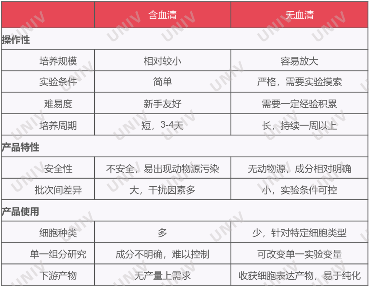
Figure 1: Differences between serum-containing and serum-free culture systems
Related Products Mentioned in this Section:
|
Catalog Number |
Name |
|
RPMI 1640 Medium (with L-Glutamine, without HEPES) |
abs9484 |
|
RPMI 1640 WITH GLUTAMINE, WITHOUT CALCIUM NITRATE |
SH30809.01 |
|
X-VIVO 15 with Gent L-Gln, Phen Red, 1 L |
04-418Q |
|
Fetal Bovine Serum (Premium) |
abs972 |
|
Australian FBS, 500ML |
SV30208.02 |
|
New Zealand FBS |
SH30406.05 |
Culture Conditions
The culture temperature for T cells is 37°C; the CO₂ concentration is typically 5%; additionally, contamination from bacteria, fungi, mycoplasma, endotoxins, viruses, etc., must be prevented.
Related Products Mentioned in this Section:
|
Product Name |
Catalog Number |
|
Penicillin-Streptomycin Solution (100×, Antibiotic-Antimycotic) |
abs9244 |
|
Mycoplasma Detection Kit (PCR method) |
abs9588 |
|
MYCOALERT DETECTION KIT- 10 TESTS |
LT07-118 |
|
Mycoplasma Elimination Reagent |
abs9375 |
|
MYCOZAP 1 TREATMENT KIT |
LT07-818 |
|
Kinetic Chromogenic Endotoxin Detection Recombinant LAL Kit |
abs50045 |
3. T Cell Activation
In a natural state, T cell activation requires two signals: The first signal is the interaction between the antigen-specific T cell receptor (TCR/CD3) on the T cell and the antigen peptide-MHC complex on the antigen-presenting cell (APC), i.e., the antigen recognition process. The first signal is essential for T cell activation but alone cannot induce T cell proliferation and cytokine release. When co-stimulatory molecules expressed on the T cell surface bind to their corresponding ligands on APCs, the second signal for T cell activation is generated. The most important of these is the binding of the CD28 molecule on T cells to the corresponding ligands B7-1 (CD80) and B7-2 (CD86) on APCs. When culturing T cells in vitro, CD3 and CD28 antibodies are used to mimic these two activation signals. Additionally, CD3 and CD28 antibodies can be coupled to magnetic beads or multimers to activate T cells, or commercially available activation/expansion kits can be used.
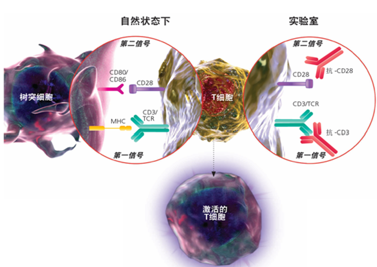
Antibody Method【3】:
a) Coat 24-well plates with anti-human CD3 antibody (diluted in sterile PBS) at a concentration of 1 μg/mL; incubate at room temperature for 4 h (or at 4°C overnight), then aspirate the unbound antibody solution;
b) Add 500 μL of PBS containing 1% BSA to each well for blocking, incubate at room temperature for 30 min, then wash once with sterile PBS;
c) Seed PBMCs into the 24-well plates at a density of 1*10⁶/mL in RPMI-1640 medium containing 10% heat-inactivated FBS, 1% penicillin/streptomycin, 3 μg/mL anti-human CD28, and 100 IU/mL human IL-2;
d) After 3 days of culture, harvest the cells, count, and assess cell viability using trypan blue staining for further analysis.
b) Add 500 μL of PBS containing 1% BSA to each well for blocking, incubate at room temperature for 30 min, then wash once with sterile PBS;
c) Seed PBMCs into the 24-well plates at a density of 1*10⁶/mL in RPMI-1640 medium containing 10% heat-inactivated FBS, 1% penicillin/streptomycin, 3 μg/mL anti-human CD28, and 100 IU/mL human IL-2;
d) After 3 days of culture, harvest the cells, count, and assess cell viability using trypan blue staining for further analysis.
Magnetic Bead Method: Taking Miltenyi Biotec's T Cell TransAct™, human as an example: humanized CD3 and CD28 antibodies are coupled to a 100 nm dextran matrix, efficiently mimicking the antigen presentation process for potent and sustained T cell activation. For use, add at a volume ratio of 1:100.
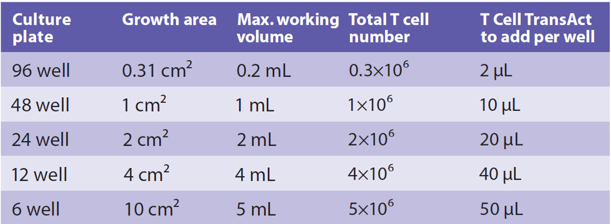
Figure 3: T cell densities and required activation reagent volumes for different plate formats
Notes:
- Primary T cells are generally cultured in vitro for no more than three weeks; for functional assays, it is recommended to use T cells cultured for less than 14 days.
- The coating concentrations of CD3 and CD28 in the antibody method need to be determined empirically, typically 1-10 ug/ml; often, CD3 is coated and CD28 is added in soluble form.
- In the magnetic bead method, beads can be micron-sized or nano-sized. For example, T Cell TransAct™, human (130-128-758, nano-sized) is removed by medium exchange or centrifugation washing, while T Cell Activation/Expansion Kit, human (130-091-441, micron-sized) uses a MACSiMAG Separator to remove beads and antibodies.
- The coating concentrations of CD3 and CD28 in the antibody method need to be determined empirically, typically 1-10 ug/ml; often, CD3 is coated and CD28 is added in soluble form.
- In the magnetic bead method, beads can be micron-sized or nano-sized. For example, T Cell TransAct™, human (130-128-758, nano-sized) is removed by medium exchange or centrifugation washing, while T Cell Activation/Expansion Kit, human (130-091-441, micron-sized) uses a MACSiMAG Separator to remove beads and antibodies.
Related Products Mentioned in this Section:
|
Product Name |
Catalog Number |
|
Invivo anti-Human CD28 Monoclonal Antibody(15E8) |
abs171614-100uL |
|
Invivo anti-Human CD3 Monoclonal Antibody(OKT3) |
abs171613-100uL |
|
T Cell Activation/Expansion kit, human |
130-091-441 |
|
T Cell Activation/Expansion Kit, mouse |
130-093-627 |
4. T Cell Expansion
T cell expansion enables researchers to generate large numbers of T cells to maximize therapeutic potential. Specific cytokines, such as interleukin-2 (IL-2) or IL-7/IL-15, are typically added to the culture to support T cell expansion and survival. A 2021 paper found that a T cell density of 2.5×10⁵ cells/mL yielded better expansion than higher or lower densities, and higher expansion efficiency was observed at IL-2 concentrations above 50 IU/mL【4】. When large numbers of antigen-specific T cells are needed, repeated antigen stimulation can be used to activate and expand T cells, but repeated activation should be used cautiously as it can lead to T cell exhaustion and reduced function. During the expansion phase, the formation of T cell clones, detection of cell proliferation, or flow cytometric analysis of corresponding cell markers can be performed.
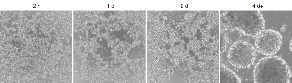
Figure 4: Clones formed during the T cell expansion phase [5]
5. T Cell Differentiation
Different types of effector T cells play central roles in the immune regulatory network, which acts as the body's defense line and triggers immune responses in disease states. For example, Th1 cytokines play important roles in recognizing and clearing intracellular pathogens such as viruses and bacteria, including Mycobacterium tuberculosis, Mycobacterium leprae, and Leishmania; Th2 cells mediate the activation and maintenance of immune responses against extracellular parasites, bacteria, allergens, and toxins; Th17 cells play a role in host defense against extracellular pathogens; Treg cells are essential for maintaining self-tolerance and immune cell homeostasis. For in vitro induction of T cell differentiation, the cytokines or antibodies required can be referenced in Figure 5. We have also compiled a summary of in vitro T cell differentiation protocols (Figure 6).

Figure 5: Naïve T cells induced to differentiate into corresponding subsets under different antibodies or cytokines
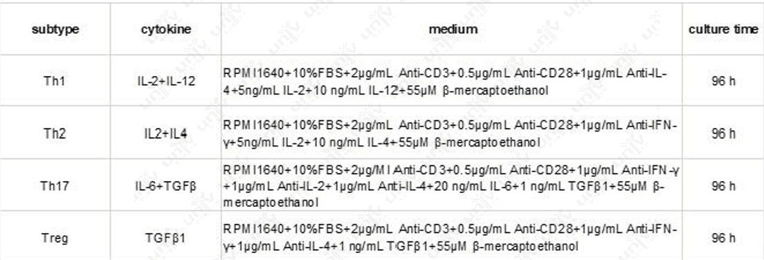
Figure 6: In vitro T cell differentiation protocols [6]
Related Products Mentioned in this Section:
|
Product Name |
Catalog Number |
|
IL-2, Human |
UA040108-500μg |
|
Recombinant Human IL-4 Protein |
abs06254 |
|
IFN-γ Protein, Human |
UA040053 |
|
Recombinant Human IL-6 Protein |
206-IL |
|
TGF-β1 Protein, Human |
UA040085-1mg |
6. T Cell Viability Maintenance
T cell viability is crucial for the success of any T cell culture experiment or therapy. Three factors need to be ensured: avoid over-confluent T cell cultures leading to density inhibition; ensure the culture system provides essential nutrients and growth factors for T cells; and maintain a sterile culture environment.
7. T Cell Functional Assays
Once a sufficient number of T cells are cultured, various experimental methods can be used to assess T cell phenotype, function, and viability, such as:
- Cytotoxicity Assays: Measure the ability of T cells to kill target cells (e.g., cancer cells or infected cells).
- ELISpot Assay: Enzyme-linked immunosorbent spot (ELISpot) assays quantify the secretion of specific cytokines, providing insight into T cell function.
- Flow Cytometry: Can be used to analyze T cell surface markers, intracellular proteins, and signal transduction.
- ELISpot Assay: Enzyme-linked immunosorbent spot (ELISpot) assays quantify the secretion of specific cytokines, providing insight into T cell function.
- Flow Cytometry: Can be used to analyze T cell surface markers, intracellular proteins, and signal transduction.

Figure 7: Surface markers, transcription factors, and secreted cytokines of different T cell subsets
Related Products Mentioned in this Section:
|
Product Name |
Catalog Number |
|
Human CAR-T Cell 9-color Panel |
LXFH09-1 |
|
Human T Cell Differentiation 12-color Panel |
LXFH12-1 |
|
Mouse Th1/Th2/Th17+Treg Cell 9-color Panel |
LXFM09-1 |
|
PE Mouse Anti-Human CD3(UCHT1) |
561880 |
|
Hu CD4 BUV661 M-T477 50ug |
750174 |
8. Hot Topics in T Cell Research
Taking the tumor organoid-T cell co-culture system described in the Nature Protocols paper "Tumor organoid--T-cell coculture systems" as an example:
Organoid part: Before starting the co-culture, tumor cells must be obtained from tumor tissues to establish tumor organoids. Two days before co-culture, tumor organoids are isolated from Matrigel. One day before co-culture, they are stimulated with IFN-γ. On the day of co-culture, tumor organoids are dissociated into single cells.
T Cell part: Isolate PBMCs from peripheral blood.
Co-culture part: Co-culture the tumor organoid single-cell suspension with PBMCs in anti-CD28 coated dishes, supplemented with IL-2 and anti-PD-1. After one week of co-culture, freshly dissociated tumor organoid single-cell suspensions are used to re-stimulate the PBMCs. After two weeks of co-culture, T cells can be cryopreserved (yellow), assessed for reactivity against tumor cells (green), or used in tumor organoid killing assays (blue).
T Cell part: Isolate PBMCs from peripheral blood.
Co-culture part: Co-culture the tumor organoid single-cell suspension with PBMCs in anti-CD28 coated dishes, supplemented with IL-2 and anti-PD-1. After one week of co-culture, freshly dissociated tumor organoid single-cell suspensions are used to re-stimulate the PBMCs. After two weeks of co-culture, T cells can be cryopreserved (yellow), assessed for reactivity against tumor cells (green), or used in tumor organoid killing assays (blue).
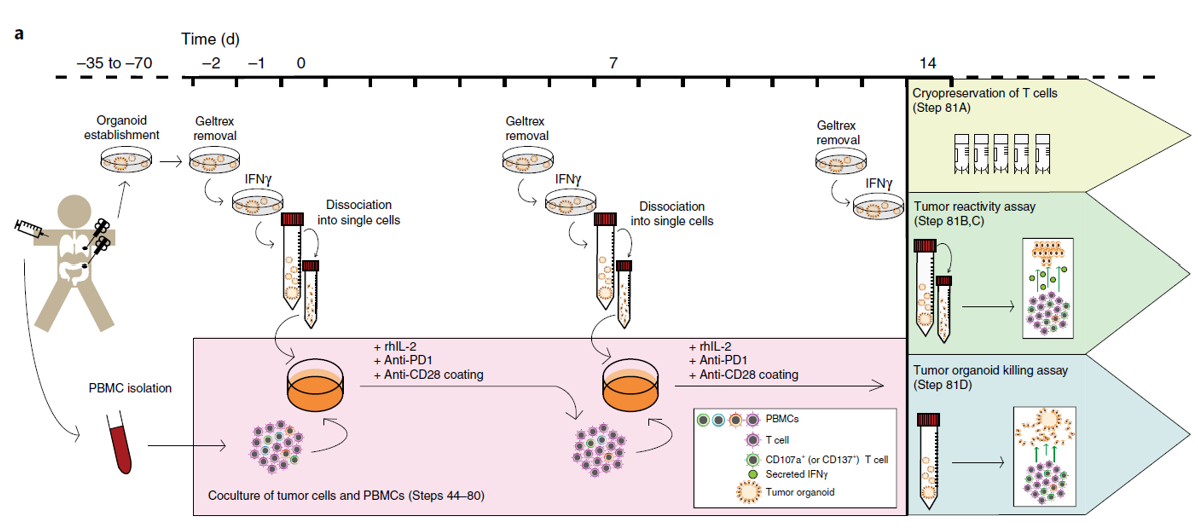
Figure 8: Flowchart of the tumor organoid-T cell co-culture system [7]
Related Products Mentioned in this Section:
|
Product Name |
Catalog Number |
|
Organotial Human Breast Cancer Organoid Culture Kit |
abs9446-1kit |
Organotial Mouse Normal Lung Organoid Culture Kit |
abs9515-1kit |
Cultrex Reduced Growth Factor Basement Membrane Extract, Type 2, Pathclear |
3533-010-02 |
|
Serum-Free Cell Cryopreservation Medium (Research Grade) |
abs9417 |
|
CryoMACS® Freezing Bags |
200-074-401 |
Notes:
- The purpose of stimulating tumor organoids with IFN-γ before co-culture is to maximize antigen presentation during the subsequent co-culture.
- Anti-PD-1 is added during co-culture to counteract the induction of PD-L1 by IFN-γ.
- When co-culturing tumor organoids with immune cells, precise control of immune cell ratios and activation status is required.
- The tumor's response to the immune system is dynamic, involving processes such as immune surveillance, immune evasion, and tumor immunoediting, all of which need to be effectively modeled.
- Anti-PD-1 is added during co-culture to counteract the induction of PD-L1 by IFN-γ.
- When co-culturing tumor organoids with immune cells, precise control of immune cell ratios and activation status is required.
- The tumor's response to the immune system is dynamic, involving processes such as immune surveillance, immune evasion, and tumor immunoediting, all of which need to be effectively modeled.
Beyond T Cells, Which Other Cells Would You Like to Learn About?
Besides the immune defenders - T cells, many other cells in the body are "working hard." There are four main tissue types in the human body: nervous tissue, connective tissue, epithelial tissue, and muscle tissue. Each cell type has its own value and research significance: neuronal activity is closely related to mood regulation and mental health; studying neurons helps understand the causes of psychological disorders like depression and anxiety; the interaction between fibroblasts and tumor cells affects tumor growth and metastasis; understanding these interactions aids in developing anti-cancer strategies; macrophages, as key cells of innate immunity, participate in various physiological and pathological processes including inflammation, tumor immunity, and autoimmune diseases.
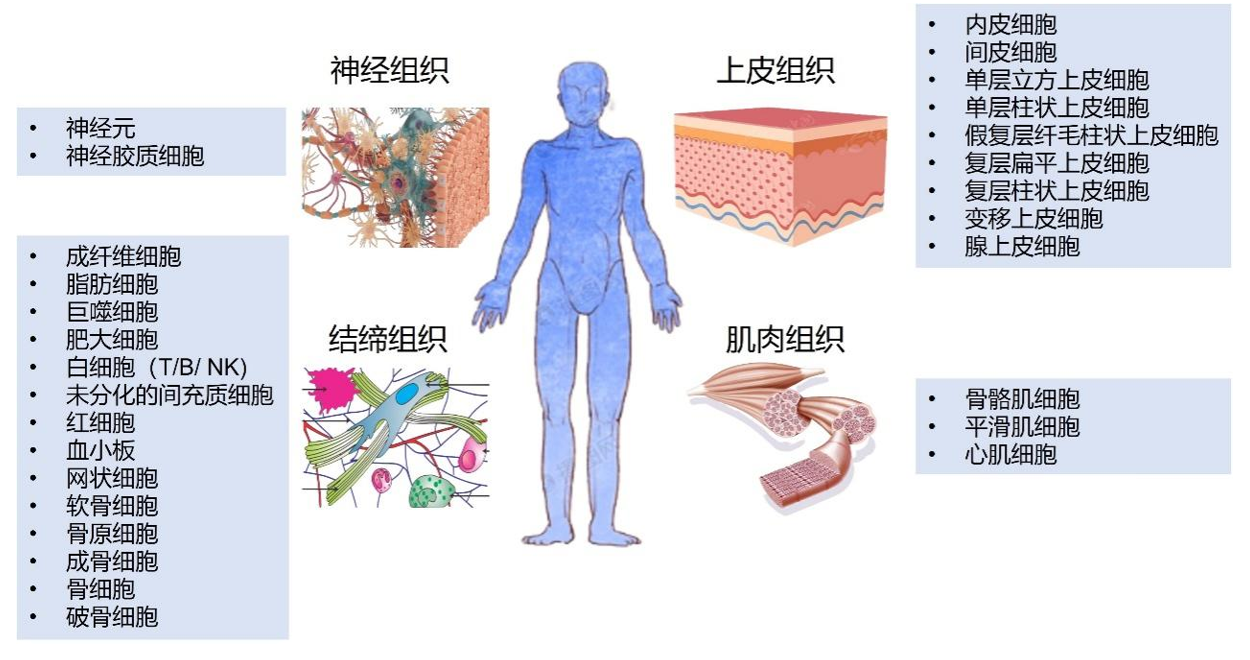 Figure 9: The four major tissue types and their corresponding cell types
For more cell types you wish to learn about, please contact your nearest UniV sales representative or the assistant mentioned below. We'll arrange it!
References:
1. Dekkers, J.F., et al., Uncovering the mode of action of engineered T cells in patient cancer organoids. Nature Biotechnology, 2022. 41(1): p. 60-69.
2. Zhou, Z., et al., A T Cell‐Engaging Tumor Organoid Platform for Pancreatic Cancer Immunotherapy. Advanced Science, 2023. 10(23).
3. Soltantoye, T., et al., Soluble and Immobilized Anti-CD3/28 Distinctively Expand and Differentiate Primary Human T Cells: An Implication for Adoptive T Cell Therapy. Iranian Journal of Allergy, Asthma and Immunology, 2022.
4. Ghaffari, S., et al., Optimizing interleukin-2 concentration, seeding density and bead-to-cell ratio of T-cell expansion for adoptive immunotherapy. BMC Immunology, 2021. 22(1).
5. Zhang, D.K.Y., A.S. Cheung, and D.J. Mooney, Activation and expansion of human T cells using artificial antigen-presenting cell scaffolds. Nature Protocols, 2020. 15(3): p. 773-798.
6. Sekiya, T. and A. Yoshimura, In Vitro Th Differentiation Protocol, in TGF-β Signaling. 2016. p. 183-191.
7. Cattaneo, C.M., et al., Tumor organoid--T-cell coculture systems. Nature Protocols, 2019. 15(1): p. 15-39.
1. Dekkers, J.F., et al., Uncovering the mode of action of engineered T cells in patient cancer organoids. Nature Biotechnology, 2022. 41(1): p. 60-69.
2. Zhou, Z., et al., A T Cell‐Engaging Tumor Organoid Platform for Pancreatic Cancer Immunotherapy. Advanced Science, 2023. 10(23).
3. Soltantoye, T., et al., Soluble and Immobilized Anti-CD3/28 Distinctively Expand and Differentiate Primary Human T Cells: An Implication for Adoptive T Cell Therapy. Iranian Journal of Allergy, Asthma and Immunology, 2022.
4. Ghaffari, S., et al., Optimizing interleukin-2 concentration, seeding density and bead-to-cell ratio of T-cell expansion for adoptive immunotherapy. BMC Immunology, 2021. 22(1).
5. Zhang, D.K.Y., A.S. Cheung, and D.J. Mooney, Activation and expansion of human T cells using artificial antigen-presenting cell scaffolds. Nature Protocols, 2020. 15(3): p. 773-798.
6. Sekiya, T. and A. Yoshimura, In Vitro Th Differentiation Protocol, in TGF-β Signaling. 2016. p. 183-191.
7. Cattaneo, C.M., et al., Tumor organoid--T-cell coculture systems. Nature Protocols, 2019. 15(1): p. 15-39.
Disclaimer: This article partially utilizes artificial intelligence assistance in its creation. If any content involves copyright or intellectual property issues, please let us know and we promise to verify and remove it as soon as possible.
Purchase recombinant protein, choose Nanjing UA-Bio
UA protein focuses on providing various protein reagents, raw materials, and services required for drug research and development, cell therapy, gene therapy, and basic scientific research, including drug target proteins, immune checkpoint proteins, cytokines, tool enzymes, customized protein expression, and full-length transmembrane protein development. Youai is committed to providing customers with high-quality products and professional services, and building a high-tech enterprise with international competitiveness.
Target proteins | membrane proteins | cytokines | enzymes | viral antigens | protein customization
Nanjing UA Biotechnology Co., Ltd. Email:order@ua-bio.com Phone:0571-87565022


Link:





©2021-2026 all rights reserved UA Biotechnology Co.,Ltd.

网站备案号 浙ICP备2022019033号-2 浙公网安备33010202004867号
 Business license (three certificates in one)
 Tel:+86-0571-87565022
E-mail:order@ua-bio.com
All products sold on this website shall not be used for clinical diagnosis or treatment of humans or animals, and shall only be used for non-medical purposes such as industrial or scientific research.
