




IL-5 standard: A core regulatory factor in eosinophil expansion and immune checkpoint therapy response
Author: Senior Product Manager of UA Views:12 


This article focuses on the molecular characteristics and biological functions of interleukin-5 (IL-5), systematically elaborating its core mechanism in immune checkpoint inhibitor therapy by driving systemic expansion of eosinophils and local tumor infiltration to enhance antitumor immune responses, analyzing the role of IL-5 in bone marrow eosinophilopoiesis and CD4+ T cell-dependent regulation, and exploring the clinical significance of the IL-5/IL-33 axis in immune cell synergy within the tumor microenvironment.
- Recent Advances
- Product Information
Recent Advances
IL-5 Standard: A Core Regulatory Factor in Eosinophil Expansion and Immune Checkpoint Therapy Response
Summary: This article focuses on the molecular characteristics and biological functions of interleukin-5 (IL-5), systematically elaborating its core mechanism in immune checkpoint inhibitor therapy by driving systemic eosinophil expansion and local tumor infiltration to enhance antitumor immune responses, analyzing the role of IL-5 in bone marrow eosinophilopoiesis and CD4+ T cell-dependent regulation, and discussing the clinical significance of the IL-5/IL-33 axis in the coordination of immune cells within the tumor microenvironment.
I. Innate Immune Cell Coordination in Immune Checkpoint Inhibitor Therapy.
Immune checkpoint inhibitors have achieved breakthrough progress in the treatment of various tumors, but the treatment response rate remains low, and the mechanisms of resistance require further exploration. Previously, the mechanisms of response to immune checkpoint inhibitors mainly focused on T cell-related immune responses; however, effective immune responses depend on the synergy between innate and adaptive immune cells, among which eosinophils are innate immune cells that have received increasing attention in recent years. Studies have reported that in melanoma, non-small cell lung cancer, and renal cell carcinoma, elevated eosinophil levels are associated with responses to anti-PD-1/PD-L1 and anti-CTLA-4 therapy. However, in tumors with low immunogenicity, whether eosinophils correlate with immunotherapy response remains unknown. In addition, the mechanisms by which eosinophils accumulate and migrate to the tumor microenvironment after immune checkpoint inhibitor treatment remain unclear. These unresolved scientific questions have driven systematic research into the role of eosinophils in immunotherapy.
II. The Core Driving Role of IL-5 in Systemic Eosinophil Expansion.
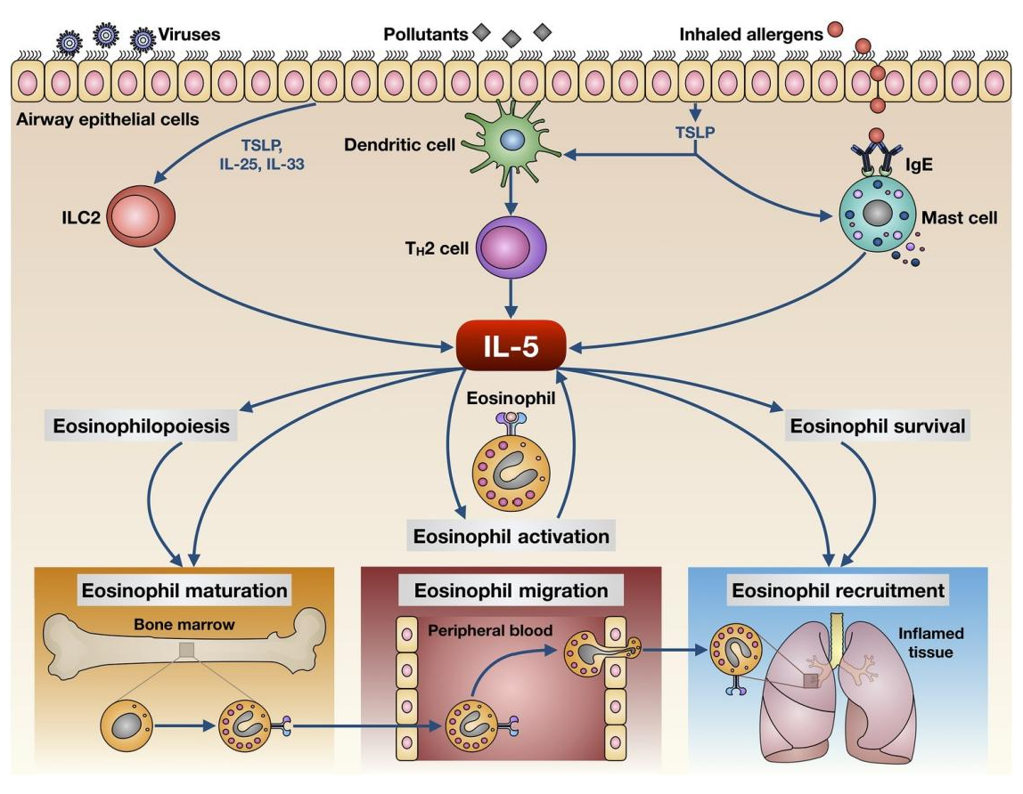
In this study, the researchers used primary and metastatic triple-negative breast cancer animal models to simulate the low immunogenicity and highly immunosuppressive characteristics of human breast cancer, observed the role played by eosinophils in the response to immune checkpoint inhibitor therapy, and explored the mechanisms of their systemic expansion and local tumor infiltration. The study found that immune checkpoint inhibitors increase IL-5 produced by CD4+ T cells, stimulating the bone marrow to produce more eosinophils, leading to systemic eosinophil expansion.
To explore the mechanism of systemic eosinophil elevation, the researchers examined eosinophil levels in the bone marrow and found that immune checkpoint inhibitors can upregulate eosinophils and their precursor cells, and also upregulate both immature and fully mature eosinophil levels. Further exploring the mechanism inducing eosinophil elevation, a series of cytokines in mouse plasma were measured, and IL-5 was found to be the only one significantly elevated. Using a patient-derived tumor fragment platform, the protein levels of IL-5 after tumor stimulation with immune checkpoint inhibitors were evaluated in vitro, and it was found that immune checkpoint inhibitor responders could induce IL-5 production.
In the metastatic tumor mouse model, when IL-5 was blocked with neutralizing antibodies, the number of eosinophils in the bone marrow, peripheral blood, and lung tissue decreased significantly, indicating that this process is IL-5-dependent. In the primary tumor mouse model, cisplatin combined with immune checkpoint inhibitors could induce elevated plasma IL-5 levels, and IL-5 neutralizing antibodies could likewise downregulate eosinophil levels in peripheral blood and tumor tissue, and could counteract the tumor-suppressive effect of cisplatin combined with immune checkpoint inhibitors. These results established the core driving position of IL-5 in immune checkpoint inhibitor-induced eosinophil expansion.
III. Cellular Sources of IL-5 and CD4+ T Cell-Dependent Regulation.
To further explore which immune cells are the source of IL-5, a rag-1 knockout mouse model was used, suggesting that adaptive immune cells are the source of IL-5 induced by immune checkpoint inhibitors. In the metastatic tumor model, immune checkpoint inhibitors could induce CD4+ CD25- T cells to produce IL-5. Using neutralizing antibodies to deplete CD4+ T cells could downregulate IL-5 expression and inhibit the immune checkpoint inhibitor-induced elevation of eosinophil levels. After treatment of patient peripheral blood mononuclear cells with immune checkpoint inhibitors, IL-5 expression increased, and the number of IL-5-producing CD4+ T cells increased. These results indicate that CD4+ T cells are the main source of immune checkpoint inhibitor-induced IL-5 production, and IL-5 further drives the generation and release of bone marrow eosinophils.
IV. Synergistic Action of IL-5 and IL-33 in the Tumor Microenvironment.
Although immune checkpoint inhibitors alone can promote systemic eosinophil upregulation, in the tumor microenvironment, only cisplatin combined with immune checkpoint inhibitors could promote eosinophil infiltration and produce effective tumor-suppressive effects. To further investigate the mechanism, relevant cytokines were screened in plasma samples from mouse models, and IL-33 was found to be significantly elevated in the cisplatin combined with immune checkpoint inhibitor group, whereas either alone could not significantly induce its elevated expression. Moreover, in clinical triple-negative breast cancer specimens, it was also observed that IL-33 expression in metastatic lesions of immune checkpoint inhibitor responders correlated with eosinophil signatures.
Using IL-33 antagonists, it was observed that the recruitment effect of IL-33 on eosinophils was limited to the tumor microenvironment and did not change peripheral blood eosinophil levels. IL-33 antagonists could likewise block the activation of CD8+ T cells by cisplatin combined with immune checkpoint inhibitors and counteract their tumor-suppressive effects, consistent with the effect of depleting eosinophils. In the metastatic tumor mouse model, recombinant IL-33 could upregulate eosinophil levels in peripheral blood, bone marrow, and tumors. Only recombinant IL-33 combined with immune checkpoint inhibitors could promote CD8+ T cell activation, promote the upregulation of the proportion of effector CD8+ T cells, and enhance tumor-suppressive effects and prolong survival. These findings revealed the synergistic action between IL-5-mediated systemic eosinophil expansion and IL-33-mediated local tumor infiltration.
V. Clinical Relevance of IL-5 in Immunotherapy Response.
By comparing patient blood samples before and after immune checkpoint inhibitor treatment, three immune cell populations associated with immune checkpoint inhibitor response were identified, namely CD1c+ dendritic cells, regulatory T cells, and eosinophils. The latter two populations were specifically upregulated in patients responding to immune checkpoint inhibitors, while CD1c+ dendritic cells were downregulated in non-responders. Eosinophil levels correlated with immunotherapy efficacy, and elevated peripheral blood eosinophil levels were associated with longer progression-free survival and overall survival. In addition, similar phenomena were observed in non-small cell lung cancer and early-stage mismatch repair-proficient colorectal cancer patients, while no significant difference was observed in mismatch repair-deficient colorectal cancer. By detecting eosinophil markers, it was found that eosinophils had higher aggregation in tumors of immune checkpoint inhibitor responders. Further analysis showed that elevated intratumoral eosinophil levels may correlate with the infiltration and activation of CD8+ T cells in the tumor microenvironment.
VI. Conclusion.
In IL-5-related basic research and drug development, high-quality recombinant IL-5 standards are core tools for dissecting its signal transduction mechanisms and evaluating its biological functions. In response to the needs of IL-5 function research and detection method development, UIA provides IL-5 Protein, Mouse. This product is suitable for applications such as mouse IL-5/IL-5R signaling pathway mechanism research, eosinophil differentiation and function research, tumor immune microenvironment analysis, and the establishment and validation of IL-5 immunoassays.
As a core driving factor of systemic eosinophil expansion in immune checkpoint inhibitor therapy, IL-5, with its unique mechanism of being produced by CD4+ T cells and stimulating bone marrow eosinophil production, as well as its synergistic action with peripheral IL-33-mediated local tumor infiltration, has become an important molecular bridge connecting adaptive immune responses and innate immune effector cells. From immune checkpoint inhibitor-induced IL-5 production by CD4+ T cells, to bone marrow eosinophil expansion, to IL-33-mediated tumor microenvironment infiltration and enhanced CD8+ T cell activation, this complete signaling axis provides a new perspective for understanding the mechanisms of immunotherapy response. IL-5 Protein, Mouse provides reliable tool support for the in-depth analysis of the IL-5 signaling network and the development of tumor immunotherapy strategies, and will continue to drive innovative exploration in the field of tumor immunology.
This article is reviewed and published by the technical expert team of UA
Disclaimer: This article partially utilizes artificial intelligence assistance in its creation. If any content involves copyright or intellectual property issues, please let us know and we promise to verify and remove it as soon as possible.
Purchase recombinant protein, choose 
UA protein focuses on providing various protein reagents, raw materials, and services required for drug research and development, cell therapy, gene therapy, and basic scientific research, including drug target proteins, immune checkpoint proteins, cytokines, tool enzymes, customized protein expression, and full-length transmembrane protein development. Nanjing Univ is committed to providing customers with high-quality products and professional services, and building a High-tech Biological Enterprise with International Competitiveness.
Target proteins | membrane proteins | cytokines | enzymes | viral antigens | protein customization
Nanjing UNIV Biotechnology Co., Ltd. Email:order@ua-bio.com Phone:+86-25-56221161

Product Information

Link:





©2021-2026 all rights reserved UNIV Biotechnology Co.,Ltd.
Privacy Policy
网站备案号 苏ICP备2025207586号-2 浙公网安备33010202004867号
 Business license (three certificates in one)
 Tel:+86-25-56221161
E-mail:order@ua-bio.com
All products sold on this website shall not be used for clinical diagnosis or treatment of humans or animals, and shall only be used for non-medical purposes such as industrial or scientific research.


