




Tetramer Staining: A Precision Technology Platform for Antigen-Specific T Cell Detection
Author: Senior Product Manager of UA Views:7 


This article focuses on the principles of tetramer staining technology and its applications in immunological research, systematically elucidating the molecular mechanism by which tetramer staining enhances TCR binding affinity through multivalent effects. It analyzes the core technical advantages of this method in the quantitative detection, sorting, and functional analysis of antigen-specific T cells, and explores its application strategies in viral infection studies, vaccine evaluation, and tumor immunology.
- Recent Advances
- Product Information
Recent Advances
Tetramer Staining: A Precision Technology Platform for Antigen-Specific T Cell Detection
Overview
This article systematically elaborates on the molecular mechanism of tetramer staining technology and its applications in immunological research. It highlights how tetramer staining enhances TCR binding affinity through multivalent effects, analyzes its core technical advantages in quantitative detection, sorting, and functional analysis of antigen-specific T cells, and explores its strategic applications in viral infection studies, vaccine evaluation, and tumor immunology.
This article systematically elaborates on the molecular mechanism of tetramer staining technology and its applications in immunological research. It highlights how tetramer staining enhances TCR binding affinity through multivalent effects, analyzes its core technical advantages in quantitative detection, sorting, and functional analysis of antigen-specific T cells, and explores its strategic applications in viral infection studies, vaccine evaluation, and tumor immunology.
I. Molecular Principles of Tetramer Staining Technology
Tetramer staining is a core technology for directly detecting antigen-specific T cells based on MHC-peptide tetramer complexes. Its fundamental principle is rooted in the immunological basis of T cell recognition of MHC-peptide complexes via TCR. However, the natural affinity between TCR and a single MHC-peptide monomer is low, with rapid dissociation rates, making stable labeling challenging. Tetramer staining technology addresses this bottleneck by using a biotin-streptavidin system to assemble four biotinylated MHC-peptide monomers with fluorescently labeled streptavidin into a tetramer complex. This multivalent effect allows a single tetramer molecule to simultaneously bind multiple TCRs on the same T cell surface, significantly slowing the overall dissociation rate by increasing local effective concentration. This ensures the stability of the labeled complex during the washing steps of flow cytometry. This ingenious molecular design makes tetramer staining a reliable tool for directly detecting antigen-specific T cells.
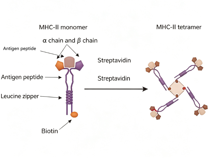
II. Technical Advantages and Workflow of Tetramer Staining
Tetramer staining offers multiple technical advantages over traditional T cell detection methods. In terms of high specificity, the technology directly identifies TCRs on antigen peptide-specific CD8+ T cells and is internationally recognized as the gold standard for antigen-specific T cell detection. In terms of high sensitivity, tetramers exhibit strong TCR affinity and low background noise, enabling the detection of rare antigen-specific T cells with frequencies as low as 0.01% to 0.1%. In terms of efficiency, the technology eliminates the need for in vitro peptide stimulation or expansion, allowing detection to be completed within 3 to 4 hours when combined with flow cytometry. In terms of quantitative accuracy, tetramer staining avoids pre-processing steps such as cell stimulation or expansion, enabling direct T cell detection and minimizing bias introduced by in vitro manipulations.
The standard workflow of tetramer staining includes four main steps: sample preparation, tetramer staining, flow cytometry analysis, and data processing. During sample preparation, heparin-anticoagulated whole blood or peripheral blood mononuclear cells are used as detection samples. In the staining step, fluorescently labeled MHC tetramers are incubated with the sample under dark conditions at room temperature, alongside lineage markers such as anti-CD3, CD4, and CD8 antibodies. A "surface staining-fixation-lysis" protocol is employed, where fixation and red blood cell lysis are performed post-staining before flow cytometry analysis. For data analysis, a gating strategy is typically applied: first, the lymphocyte population is identified, followed by the CD3+CD8+ T cell subset, and finally, the proportion and fluorescence intensity of tetramer-positive cells within the CD8+ T cell population are analyzed.
III. Applications of Tetramer Staining in Viral Infection Research
Tetramer staining plays a central role in viral infection research. In the detection of virus-specific T cell immunity, the technology can be used to quantitatively analyze the frequency and dynamic changes of antigen-specific CD8+ T cells in peripheral blood or tissues post-infection. For example, in Epstein-Barr virus (EBV) infection, the host mounts a robust virus-specific T cell response. Using EBV LMP1-specific tetramer staining, the expansion and memory formation of virus-specific T cells can be precisely monitored. In mechanistic studies, combining tetramer staining with memory and functional marker staining enables in-depth analysis of the differentiation trajectory and functional states of virus-specific T cells. In transplantation medicine, tetramer staining can monitor the immune reconstitution status of donor-derived common virus-specific (e.g., CMV, EBV) T cells, providing a basis for adjusting post-transplant immunosuppressive regimens.
IV. Applications of Tetramer Staining in Vaccine Evaluation and Tumor Immunology
In vaccine development, tetramer staining can be used to screen antigen epitopes for vaccine design. By detecting the strength of T cell responses induced by different candidate epitopes, the most immunodominant epitopes can be selected for vaccine formulation. In vaccine efficacy monitoring, the technology can assess the extent and persistence of antigen-specific T cell expansion post-vaccination. In tumor immunology research, tetramer staining aids in identifying tumor-specific antigens, detecting the presence and frequency of antigen-specific T cells among tumor-infiltrating lymphocytes, and evaluating tumor immunogenicity and potential patient responses to immunotherapy. In cell therapy, the technology facilitates quality control of CAR-T and TCR-T cell products by enabling the sorting of antigen-specific T cells for in vitro expansion and functional analysis.
V. Conclusion
In practical applications of tetramer staining, high-quality MHC tetramer reagents are critical for ensuring detection specificity and data reliability. To meet the needs of EBV-related immunological research, UA offers UA-MHC HLA-A*0201/YLLEILWRL EBV LMP1 Tetramer-PE Labelled. This product is a PE-labeled tetramer complex that specifically identifies HLA-A0201-restricted EBV LMP1 epitope-specific CD8+ T cells. It is suitable for applications such as EBV infection immune monitoring, EBV-specific T cell frequency analysis, and post-transplant immune reconstitution assessment.
Tetramer staining technology, with its ingenious molecular design that enhances TCR binding affinity through multivalent effects and its high compatibility with flow cytometry platforms, has become the gold standard method for antigen-specific T cell detection. From viral infection immune monitoring to vaccine immunogenicity evaluation, from tumor-specific antigen screening to cell therapy product quality control, tetramer staining continues to play an indispensable role in both basic immunological research and clinical translation. UA-MHC HLA-A*0201/YLLEILWRL EBV LMP1 Tetramer-PE Labelled provides a reliable tool for detecting and monitoring EBV-specific T cells, driving further exploration in the field of viral infection immunology.
This article is reviewed and published by the technical expert team of UA
Disclaimer: This article partially utilizes artificial intelligence assistance in its creation. If any content involves copyright or intellectual property issues, please let us know and we promise to verify and remove it as soon as possible.
Purchase recombinant protein, choose Nanjing UA-Bio
UA protein focuses on providing various protein reagents, raw materials, and services required for drug research and development, cell therapy, gene therapy, and basic scientific research, including drug target proteins, immune checkpoint proteins, cytokines, tool enzymes, customized protein expression, and full-length transmembrane protein development. Youai is committed to providing customers with high-quality products and professional services, and building a High-tech Biological Enterprise with International Competitiveness.
Target proteins | membrane proteins | cytokines | enzymes | viral antigens | protein customization
Nanjing UA Biotechnology Co., Ltd. Email:order@ua-bio.com Phone:+86-25-56221161

Product Information

Link:





©2021-2026 all rights reserved UA Biotechnology Co.,Ltd.
Privacy Policy 
网站备案号 浙ICP备2022019033号-2 浙公网安备33010202004867号
 Business license (three certificates in one)
 Tel:+86-25-56221161
E-mail:order@ua-bio.com
All products sold on this website shall not be used for clinical diagnosis or treatment of humans or animals, and shall only be used for non-medical purposes such as industrial or scientific research.


