




CCL1: Key regulatory factors of Treg cell-driven macrophage efferocytosis and tissue repair
Author: Senior Product Manager of UA Views:7 


This article systematically elucidates the molecular characteristics and biological functions of the chemokine CCL1, focusing on its role as a key effector secreted by regulatory T cells to enhance macrophage efferocytosis and construct a pro-regenerative immune microenvironment through the STAT3–SCARB1 signaling pathway. It further analyzes its dual tissue-repair functions in skin wound healing and colonic mucosal repair.
- Recent Advances
- Product Information
Recent Advances
CCL1: A Key Regulator of Treg Cell-Driven Macrophage Efferocytosis and Tissue Repair
Overview
This article systematically elaborates on the molecular characteristics and biological functions of the chemokine CCL1, highlighting its role as a key effector secreted by regulatory T cells (Tregs). It explores how CCL1 enhances macrophage efferocytosis and constructs a pro-regenerative immune microenvironment through the STAT3-SCARB1 signaling pathway, while analyzing its dual tissue repair functions in skin wound healing and colonic mucosal repair.
This article systematically elaborates on the molecular characteristics and biological functions of the chemokine CCL1, highlighting its role as a key effector secreted by regulatory T cells (Tregs). It explores how CCL1 enhances macrophage efferocytosis and constructs a pro-regenerative immune microenvironment through the STAT3-SCARB1 signaling pathway, while analyzing its dual tissue repair functions in skin wound healing and colonic mucosal repair.
I. Molecular Characteristics and Cellular Sources of CCL1
CCL1 (C-C motif chemokine ligand 1), also known as I-309 or TCA-3, is an important member of the CC chemokine family. The human CCL1 gene is located on chromosome 17q11.2, encoding a mature protein composed of approximately 74 amino acid residues with a molecular weight of about 8.5 kDa. The primary structure of CCL1 contains four highly conserved cysteine residues, forming two intramolecular disulfide bonds (Cys10-Cys34 and Cys11-Cys35) that maintain its tertiary structure stability. CCL1 is primarily secreted by activated regulatory T cells (Tregs), but it can also be produced by activated Th2 cells, mast cells, and monocytes. The only known functional receptor for CCL1 is CCR8, a seven-transmembrane G protein-coupled receptor expressed on macrophages, Th2 cells, Tregs, and some tumor cells. The binding affinity between CCL1 and CCR8 is at the nanomolar level, activating downstream Gi protein-mediated signaling pathways to exert chemotactic and immunomodulatory functions.
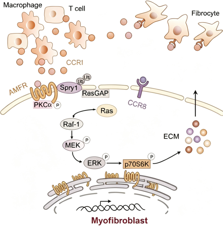
II. Immunomodulatory and Tissue Repair Functions of Treg-Derived CCL1
Regulatory T cells are a key subset of immune cells with immunosuppressive functions. Recent studies have revealed that Tregs not only mediate immune regulation through cell contact and inhibitory cytokines but also participate in tissue repair and regeneration by secreting chemokines such as CCL1. Treg-derived CCL1 enhances macrophage efferocytosis—the process by which macrophages recognize, phagocytose, and clear apoptotic cells, a critical step in inflammation resolution and tissue repair. By acting on the CCR8 receptor on macrophages, CCL1 improves their efficiency in clearing apoptotic neutrophils and epithelial cells, thereby reducing the release of pro-inflammatory contents from apoptotic cells that damage surrounding tissues and promoting macrophage polarization toward a repair-oriented (M2-like) phenotype. This process occurs simultaneously in colonic tissue and skin wounds, establishing a pro-regenerative immune microenvironment.
III. Molecular Mechanism of CCL1-Mediated Efferocytosis Enhancement via the STAT3-SCARB1 Signaling Pathway
The molecular mechanism by which CCL1 enhances macrophage efferocytosis involves the activation of the STAT3-SCARB1 signaling pathway. SCARB1 (scavenger receptor class B member 1, also known as SR-BI) is a scavenger receptor that plays a key role in macrophage recognition and phagocytosis of apoptotic cells. Studies show that after CCL1 binds to CCR8 on macrophages, it activates the JAK2-STAT3 signaling pathway. Phosphorylated STAT3 translocates to the nucleus, upregulating SCARB1 gene expression. Increased SCARB1 protein expression enhances macrophage recognition and binding of apoptotic cells, significantly improving efferocytosis efficiency. Additionally, CCL1-mediated STAT3 activation promotes macrophage secretion of anti-inflammatory cytokines such as IL-10 and TGF-β, further consolidating the establishment of a repair-oriented microenvironment. This discovery of the signaling axis reveals a new mechanism by which Tregs regulate macrophage function through chemokines.
IV. Applications of CCL1 in Skin Wound Healing and Colonic Mucosal Repair
CCL1 exhibits dual functions in tissue repair. In skin wound healing, Tregs accumulate at the injury site and secrete CCL1, which enhances macrophage clearance of apoptotic neutrophils, reducing the duration of neutrophil infiltration and promoting the transition from inflammation to repair. This accelerates re-epithelialization and granulation tissue formation. In colonic mucosal repair, CCL1 enhances macrophage clearance of apoptotic epithelial cells via the STAT3-SCARB1 pathway, reducing mucosal inflammatory responses and promoting epithelial barrier repair and regeneration. In models of inflammatory bowel diseases such as ulcerative colitis and Crohn's disease, CCL1-mediated enhancement of macrophage efferocytosis is closely associated with accelerated mucosal healing. The discovery of this dual tissue repair function provides a theoretical basis for developing CCL1-based pro-repair therapeutic strategies.
V. Conclusion
As a key effector secreted by Tregs, CCL1 plays an irreplaceable central role in skin wound healing and colonic mucosal repair through its unique ability to enhance macrophage efferocytosis and construct a repair-oriented immune microenvironment via the STAT3-SCARB1 signaling pathway. This discovery not only reveals a new mechanism by which Tregs regulate tissue regeneration but also provides an important theoretical foundation for developing chemokine-based pro-repair therapeutic strategies. CCL1 Protein, Human serves as a critical tool for in-depth analysis of the CCL1/CCR8 signaling axis and its functional network in tissue repair.
In CCL1-related basic research and drug development, high-quality human recombinant CCL1 protein is a core tool for receptor binding analysis, macrophage functional studies, and signaling pathway exploration. To meet this research demand, Uni offers CCL1 Protein, Human, suitable for applications such as CCL1/CCR8 binding activity analysis, STAT3-SCARB1 signaling pathway mechanism exploration, and macrophage efferocytosis studies.
This article is reviewed and published by the technical expert team of UA
Disclaimer: This article partially utilizes artificial intelligence assistance in its creation. If any content involves copyright or intellectual property issues, please let us know and we promise to verify and remove it as soon as possible.
Purchase recombinant protein, choose Nanjing UA-Bio
UA protein focuses on providing various protein reagents, raw materials, and services required for drug research and development, cell therapy, gene therapy, and basic scientific research, including drug target proteins, immune checkpoint proteins, cytokines, tool enzymes, customized protein expression, and full-length transmembrane protein development. Youai is committed to providing customers with high-quality products and professional services, and building a High-tech Biological Enterprise with International Competitiveness.
Target proteins | membrane proteins | cytokines | enzymes | viral antigens | protein customization
Nanjing UA Biotechnology Co., Ltd. Email:order@ua-bio.com Phone:+86-25-56221161

Product Information

Link:





©2021-2026 all rights reserved UA Biotechnology Co.,Ltd.
Privacy Policy 
网站备案号 浙ICP备2022019033号-2 浙公网安备33010202004867号
 Business license (three certificates in one)
 Tel:+86-25-56221161
E-mail:order@ua-bio.com
All products sold on this website shall not be used for clinical diagnosis or treatment of humans or animals, and shall only be used for non-medical purposes such as industrial or scientific research.
