




Annexin V: The Gold Standard Probe for Apoptosis Detection and Molecular Recognition Tool for Phosphatidylserine Exposure
Author: Senior Product Manager of UA Views:1 


This article systematically elaborates on the molecular characteristics and biological functions of Annexin V protein, focusing on its high-affinity recognition mechanism for phosphatidylserine as a calcium-dependent phospholipid-binding protein. It analyzes its sensitivity advantages in early apoptosis detection compared to other methods and discusses the interpretation standards and experimental considerations of the Annexin V/PI double-staining method in flow cytometry.
- Recent Advances
- Product Information
Recent Advances
Annexin V: The Gold Standard Probe for Apoptosis Detection and Molecular Recognition Tool for Phosphatidylserine Exposure
Summary
This article systematically elaborates on the molecular characteristics and biological functions of Annexin V protein, focusing on its high-affinity recognition mechanism for phosphatidylserine (PS) as a calcium-dependent phospholipid-binding protein. It analyzes its sensitivity advantages in early apoptosis detection compared to other methods and discusses the interpretation standards and experimental considerations of Annexin V/PI dual staining in flow cytometry.
This article systematically elaborates on the molecular characteristics and biological functions of Annexin V protein, focusing on its high-affinity recognition mechanism for phosphatidylserine (PS) as a calcium-dependent phospholipid-binding protein. It analyzes its sensitivity advantages in early apoptosis detection compared to other methods and discusses the interpretation standards and experimental considerations of Annexin V/PI dual staining in flow cytometry.
I. Molecular Characteristics and Phospholipid-Binding Properties of Annexin V
Annexin V, a key member of the Annexin family, is a calcium-dependent phospholipid-binding protein with a molecular weight of approximately 35–36 kDa. Composed of 319 amino acid residues, its molecular structure features four homologous domains, each consisting of five α-helices, forming a characteristic disk-like conformation. In the presence of calcium ions, the four domains of Annexin V cooperatively form a high-affinity binding site for phosphatidylserine (PS), with binding affinity at the nanomolar level. Annexin V is widely distributed in various human tissues, particularly abundant in placental and vascular endothelial cells, and its physiological functions are closely related to membrane repair, blood coagulation regulation, and inflammatory response modulation. The high selectivity of Annexin V for PS stems from the precise coordination between its unique calcium-binding sites and the phosphate groups of PS—when calcium ions bind to Annexin V, the protein undergoes conformational changes, exposing a hydrophobic surface that enables high-specificity recognition of PS.
II. Phosphatidylserine Exposure as an Early Marker of Apoptosis
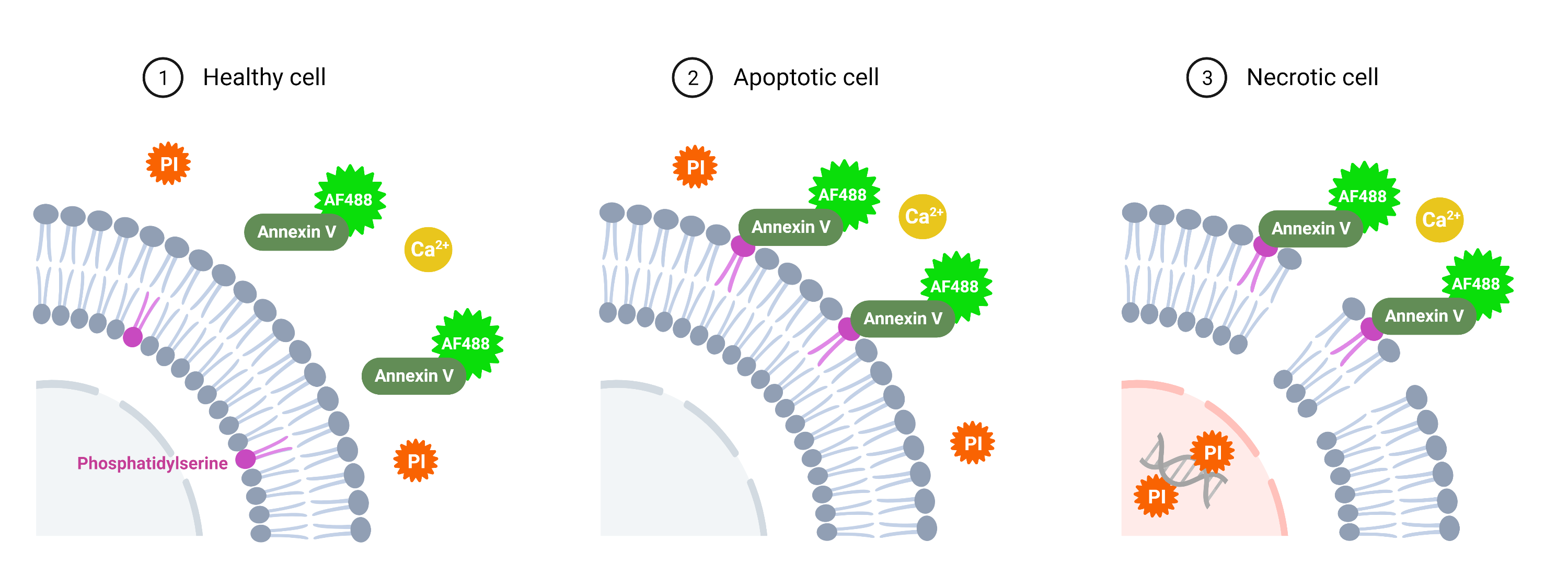
In normal living cells, phosphatidylserine (PS) is strictly distributed on the inner (cytoplasmic) leaflet of the cell membrane, maintained by the coordinated activity of ATP-dependent flippases and scramblases. When apoptosis occurs, decreased ATP levels lead to reduced flippase activity, while scramblases are activated, causing PS to rapidly translocate from the inner to the outer (extracellular) leaflet of the cell membrane. This PS exposure event occurs during the early stages of apoptosis, preceding subsequent apoptotic events such as mitochondrial membrane potential loss, caspase activation, DNA fragmentation, and loss of membrane integrity. Therefore, PS exposure is recognized as one of the earliest and most sensitive molecular markers of apoptosis. Using Annexin V, which exhibits high affinity for PS, as a probe enables early detection of apoptotic cells. Its sensitivity is significantly superior to TUNEL assays, which rely on DNA fragmentation, and it is more convenient, time-saving, and reliable, making it one of the most ideal methods for apoptosis detection.
III. Detection Principle of Annexin V/PI Dual Staining
Since necrotic cells and late-stage apoptotic cells lose membrane integrity, PS is also exposed on the outer leaflet, and the cell membrane becomes permeable to propidium iodide (PI). Using Annexin V alone cannot distinguish apoptotic cells from necrotic cells. Therefore, labeling Annexin V with a fluorophore (e.g., fluorescein isothiocyanate, FITC) and combining it with PI exclusion staining enables effective differentiation of cells in different states. The detection principle is as follows: normal living cells exhibit low staining for both Annexin V and PI; apoptotic cells exhibit high Annexin V staining and low PI staining; necrotic cells exhibit high staining for both Annexin V and PI. Cells with damaged membranes can be stained by PI, producing red fluorescence, whereas cells with intact membranes do not produce red fluorescence signals. Thus, PI does not stain cells in the early stages of apoptosis. In bivariate flow cytometry scatter plots, the lower-left quadrant represents living cells (FITC⁻/PI⁻); the upper-right quadrant represents non-viable (necrotic) cells (FITC⁺/PI⁺); the lower-right quadrant represents apoptotic cells (FITC⁺/PI⁻); and the upper-left quadrant represents mechanically damaged cell debris (FITC⁻/PI⁺). Special attention should be paid to the fact that during the late stages of apoptosis (lysis phase), cell debris may also show significant positive staining, potentially interfering with result interpretation.
IV. Experimental Considerations and Optimization Strategies for Annexin V Detection
In Annexin V apoptosis detection experiments, the following key factors should be noted. Calcium ion concentration is critical for Annexin V binding to PS; thus, calcium-containing binding buffer should be used, and buffers containing EDTA or EGTA should be avoided. Over-digestion with trypsin during cell harvesting should be avoided to prevent membrane damage and false-positive results. PI staining time should not exceed 30 minutes before detection. For adherent cells, gentle digestion solutions without EDTA are recommended for cell collection. For suspension cells, direct centrifugation followed by staining is sufficient. Additionally, Annexin V can be used to detect other cell types with exposed PS, such as activated platelets, senescent red blood cells, and chemotherapy-induced apoptotic tumor cells.
V. Conclusion
As the gold standard probe for recognizing phosphatidylserine exposure—an early apoptotic marker—Annexin V, with its high affinity for PS, calcium-dependent binding mechanism, and precise discrimination capability in flow cytometry when combined with PI dual staining, has become an indispensable core tool in apoptosis detection. Its comprehensive advantages in early apoptosis detection sensitivity, operational convenience, and result reliability ensure its irreplaceable role in basic research and drug development. Human recombinant Annexin V protein provides critical tool support for the development of apoptosis detection reagents and apoptosis mechanism research.
In apoptosis-related basic research and drug screening, high-quality human recombinant Annexin V protein is a core tool for establishing apoptosis detection methods, studying apoptosis mechanisms, and evaluating drug activity. To meet these research needs, U-Impact offers Annexin V His Tag Protein, Human, suitable for applications such as FITC-labeled apoptosis detection reagent development, Annexin V/PS binding activity analysis, and apoptosis mechanism research.
This article is reviewed and published by the technical expert team of UA
Disclaimer: This article partially utilizes artificial intelligence assistance in its creation. If any content involves copyright or intellectual property issues, please let us know and we promise to verify and remove it as soon as possible.
Purchase recombinant protein, choose Nanjing UA-Bio
UA protein focuses on providing various protein reagents, raw materials, and services required for drug research and development, cell therapy, gene therapy, and basic scientific research, including drug target proteins, immune checkpoint proteins, cytokines, tool enzymes, customized protein expression, and full-length transmembrane protein development. Youai is committed to providing customers with high-quality products and professional services, and building a High-tech Biological Enterprise with International Competitiveness.
Target proteins | membrane proteins | cytokines | enzymes | viral antigens | protein customization
Nanjing UA Biotechnology Co., Ltd. Email:order@ua-bio.com Phone:+86-25-56221161

Product Information

Link:





©2021-2026 all rights reserved UA Biotechnology Co.,Ltd.
Privacy Policy 
网站备案号 浙ICP备2022019033号-2 浙公网安备33010202004867号
 Business license (three certificates in one)
 Tel:+86-25-56221161
E-mail:order@ua-bio.com
All products sold on this website shall not be used for clinical diagnosis or treatment of humans or animals, and shall only be used for non-medical purposes such as industrial or scientific research.
