




Protein Labeling Receptors: Key Technologies from Molecular Probes to Cellular Function Analysis
Author: Senior Product Manager of UA Views:3 


本文围绕蛋白质标记受体的技术原理与应用,系统阐述荧光标记受体蛋白在流式细胞术、受体-配体相互作用分析及细胞功能研究中的核心价值,分析其在免疫细胞亚群分析、抗体药物筛选和细胞治疗产品质量评价中的应用策略。
- Recent Advances
- Product Information
Recent Advances
Protein-Labeled Receptors: Key Technology from Molecular Probes to Cellular Function Analysis
Summary
This article systematically elaborates on the technical principles and applications of protein-labeled receptors, highlighting the core value of fluorescently labeled receptor proteins in flow cytometry, receptor-ligand interaction analysis, and cellular function studies. It analyzes their application strategies in immune cell subset analysis, antibody drug screening, and quality evaluation of cell therapy products.
This article systematically elaborates on the technical principles and applications of protein-labeled receptors, highlighting the core value of fluorescently labeled receptor proteins in flow cytometry, receptor-ligand interaction analysis, and cellular function studies. It analyzes their application strategies in immune cell subset analysis, antibody drug screening, and quality evaluation of cell therapy products.
I. Technical Basis of Protein-Labeled Receptors
Protein-labeled receptors refer to receptor proteins conjugated with reporter molecules (such as fluorescent dyes, biotin, enzymes, or radioactive isotopes) through chemical coupling or genetic engineering methods, enabling visual tracking and quantitative analysis of receptor expression, distribution, and function. In immunology research and drug development, fluorescently labeled receptor proteins are one of the most widely used tools. The core technology relies on covalent coupling reactions between fluorescent dyes and specific amino acid residues (e.g., primary amine groups of lysine or thiol groups of cysteine) on the protein surface, or indirect labeling via the biotin-streptavidin system. The labeled receptor proteins retain their natural ligand-binding activity and receptor specificity while acquiring optical signals detectable by flow cytometry or fluorescence microscopy, serving as a critical bridge connecting molecular recognition and quantitative analysis.
II. Molecular Characteristics of CD19 as a B Cell Marker
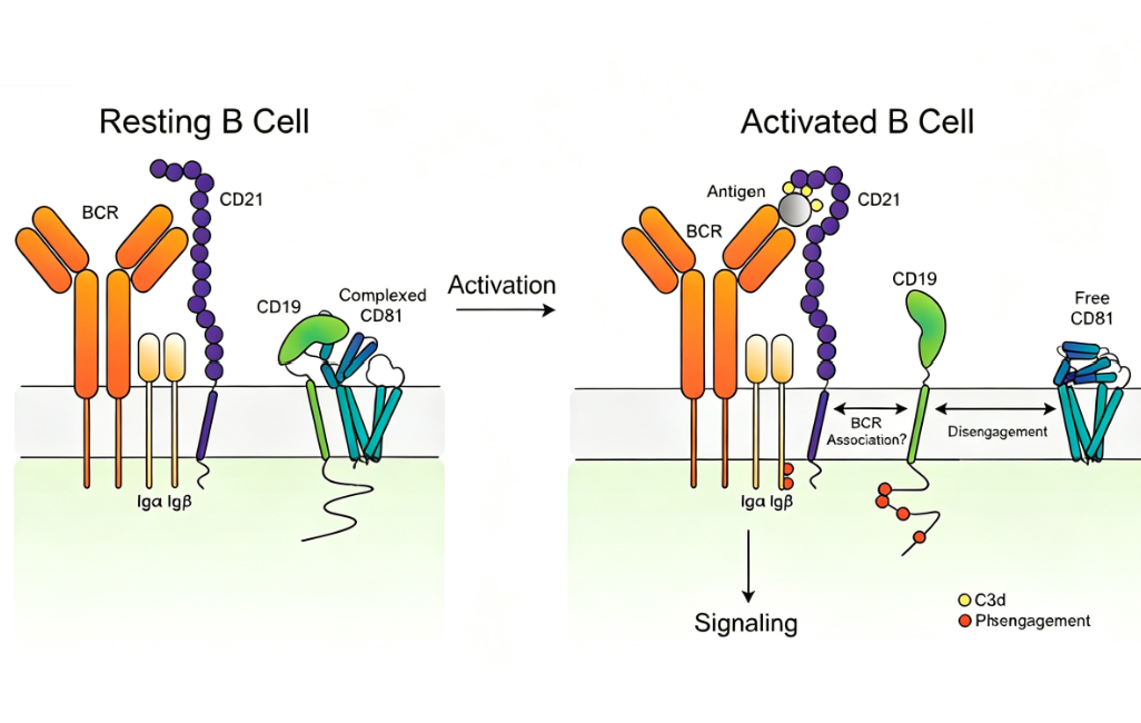
CD19 is a transmembrane glycoprotein specifically expressed in the B lymphocyte lineage and belongs to the immunoglobulin superfamily. The human CD19 gene is located on chromosome 16p11.2, encoding a protein composed of 556 amino acid residues with a molecular weight of approximately 95 kDa. Structurally, the extracellular domain of CD19 contains two C2-type immunoglobulin-like domains with multiple N-linked glycosylation sites; the transmembrane region consists of a single-pass helix; and the intracellular domain includes nine highly conserved tyrosine residues that serve as docking sites for SH2 domain-containing effector molecules upon phosphorylation. CD19 is expressed continuously from pro-B cells through pre-B cells, immature B cells, and mature B cells, until its loss upon terminal differentiation into plasma cells. All B cell lineages except plasma cells, as well as follicular dendritic cells, express this molecule. CD19 forms a B cell co-receptor complex with CD21 and CD81, enhancing BCR signal transduction to lower the antigen threshold for B cell activation, playing a central regulatory role in humoral immune responses.
III. Applications of Fluorescently Labeled Receptor Proteins in Flow Cytometry
In B cell-related basic research and clinical testing, fluorescently labeled receptor proteins are among the core tools for flow cytometry analysis. Their applications include the following aspects. In B cell subset analysis, fluorescently labeled CD19 protein can be combined with lineage markers such as anti-CD3 and anti-CD56 to achieve multicolor flow cytometry, accurately distinguishing B cells from other lymphocyte subsets. In CAR-T cell functional evaluation, the quality control of anti-CD19 CAR-T cell products relies on precise detection of CAR expression positivity and binding activity—incubating fluorescently labeled CD19 protein with CAR-T cells and detecting fluorescence signals via flow cytometry allows quantitative assessment of CAR-positive cell proportions. In antibody drug screening, labeled proteins can be used to evaluate the competition efficiency of CD19-targeting antibody drugs or bispecific antibodies for CD19 binding sites.
IV. Technical Features and Selection Criteria for Fluorescently Labeled Receptor Proteins
In practical applications of fluorescently labeled receptor proteins, the choice of fluorescent dye directly affects detection sensitivity and signal-to-noise ratio. Alexa Fluor 647, as a far-red fluorescent dye, has a maximum excitation wavelength of 650 nm and a maximum emission wavelength of 668 nm. The low cellular autofluorescence background in this wavelength range makes it suitable for flow cytometry detection systems equipped with red laser excitation sources. Additionally, the purity, endotoxin levels, and batch-to-batch consistency of labeled proteins are critical quality parameters for ensuring experimental reproducibility. His-tagged receptor proteins facilitate high-purity purification via immobilized metal affinity chromatography and allow convenient detection and capture using anti-His tag antibodies. During storage and use, labeled proteins should be strictly protected from light and avoid repeated freeze-thaw cycles to maintain fluorescence signal stability.
V. Extended Applications and Prospects of Protein-Labeled Receptor Technology
Beyond CD19, fluorescently labeled receptor protein technology has been widely applied to the detection and analysis of various immune cell markers, including CD3, CD4, CD8, CD56, and CD25. In the field of tumor immunotherapy, labeled receptor proteins can be used to monitor the expansion and persistence of CAR-T cells in vivo; in autoimmune disease research, they can analyze the frequency and phenotype of autoreactive B cells; in vaccine development, they can assess vaccine-induced antigen-specific B cell responses. With the continuous advancement of multicolor flow cytometry and spectral flow cytometry, the application scope of fluorescently labeled receptor proteins will further expand, providing more powerful tools for immunology research and clinical diagnostics.
VI. Conclusion
Protein-labeled receptor technology, as a core tool connecting molecular recognition and quantitative analysis, plays an irreplaceable role in basic immunology research, drug development, and clinical diagnostics. To meet the needs of B cell-related research and CAR-T cell product quality evaluation, U-Pharm offers Alexa Fluor 647-Labeled CD19 His Tag Protein, Human. This product features precise molecular design and excellent fluorescence labeling performance, is highly compatible with flow cytometry platforms, and provides stable and reliable detection tools for related research. With ongoing advancements in fluorescence labeling technology and detection platforms, labeled receptor proteins will play a key role in broader biomedical research fields.
This article is reviewed and published by the technical expert team of UA
Disclaimer: This article partially utilizes artificial intelligence assistance in its creation. If any content involves copyright or intellectual property issues, please let us know and we promise to verify and remove it as soon as possible.
Purchase recombinant protein, choose Nanjing UA-Bio
UA protein focuses on providing various protein reagents, raw materials, and services required for drug research and development, cell therapy, gene therapy, and basic scientific research, including drug target proteins, immune checkpoint proteins, cytokines, tool enzymes, customized protein expression, and full-length transmembrane protein development. Youai is committed to providing customers with high-quality products and professional services, and building a High-tech Biological Enterprise with International Competitiveness.
Target proteins | membrane proteins | cytokines | enzymes | viral antigens | protein customization
Nanjing UA Biotechnology Co., Ltd. Email:order@ua-bio.com Phone:+86-25-56221161

Product Information

Link:





©2021-2026 all rights reserved UA Biotechnology Co.,Ltd.
Privacy Policy 
网站备案号 浙ICP备2022019033号-2 浙公网安备33010202004867号
 Business license (three certificates in one)
 Tel:+86-25-56221161
E-mail:order@ua-bio.com
All products sold on this website shall not be used for clinical diagnosis or treatment of humans or animals, and shall only be used for non-medical purposes such as industrial or scientific research.
