




Research Tools and Application Strategies for RAW264.7 Macrophage Polarization
Author: Senior Product Manager of UA Views:9 


This article systematically elaborates on the biological characteristics of the RAW264.7 cell line as a classic model for macrophage polarization research and its standardized protocols for in vitro induction of M1/M2 polarization, analyzing the pivotal role of cytokines and TLR agonists in regulating the switch between pro-inflammatory and anti-inflammatory phenotypes.
- Recent Advances
- Product Information
Recent Advances
Research Tools and Application Strategies for RAW264.7 Macrophage Polarization
Overview
This article systematically elaborates on the biological characteristics of the RAW264.7 cell line as a classic model for macrophage polarization research and its standardized protocols for inducing M1/M2 polarization in vitro. It analyzes the pivotal role of cytokines and TLR agonists in regulating the switch between pro-inflammatory and anti-inflammatory phenotypes.
This article systematically elaborates on the biological characteristics of the RAW264.7 cell line as a classic model for macrophage polarization research and its standardized protocols for inducing M1/M2 polarization in vitro. It analyzes the pivotal role of cytokines and TLR agonists in regulating the switch between pro-inflammatory and anti-inflammatory phenotypes.
I. Advantages of the RAW264.7 Cell Line as a Macrophage Polarization Model
RAW264.7 is a murine monocyte/macrophage leukemia cell line. Due to its stable origin, ease of culture, rapid growth cycle, and clear response to stimuli, it has become the gold standard model for studying macrophage biology, immune responses, inflammatory diseases, and tumor immunology. Compared to primary macrophages isolated from tissues, RAW264.7 cells overcome limitations such as difficult acquisition, significant individual variability, and limited passage numbers. This cell line can be directionally induced to differentiate into M1-type (classically activated/pro-inflammatory) or M2-type (alternatively activated/anti-inflammatory) macrophages in vitro, with highly controllable experimental conditions and strong reproducibility. However, it is important to note that the number of passages should not be excessive—recommended to be maintained within 10 passages—to avoid spontaneous differentiation or functional drift.
II. Induction Protocol and Marker Characteristics of M1-Type Macrophages
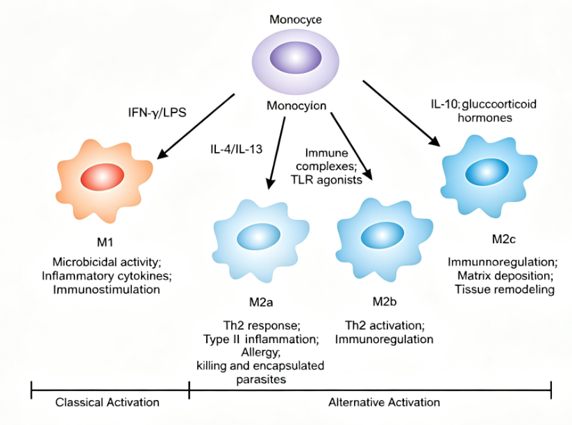
M1-type macrophages play a key role in inflammatory responses and anti-tumor immunity, secreting large amounts of pro-inflammatory cytokines such as TNF-α, IL-6, and IL-12, which promote inflammatory responses and cytotoxic effects. The classic induction protocol for polarizing RAW264.7 cells into M1-type involves the combined use of LPS (lipopolysaccharide) and IFN-γ (interferon-γ), which synergistically activate the TLR4/NF-κB and JAK/STAT signaling pathways. In standard experimental procedures, RAW264.7 cells in the logarithmic growth phase are seeded at a density of 2×10⁵ to 3×10⁵ cells per well in a six-well plate. After overnight adherence, the medium is replaced with complete medium containing 100 ng/mL LPS and 20 ng/mL IFN-γ, and cultured at 37°C with 5% CO₂ for 24 to 48 hours. Induced M1 cells exhibit morphological changes from round to irregular polygonal or amoeboid shapes, with numerous filopodia and lamellipodia extending from the edges and increased cytoplasmic granules. M1 polarization can be verified by flow cytometry to detect upregulated expression of M1 markers CD86 and MHC-II, ELISA to measure elevated levels of pro-inflammatory factors TNF-α, IL-1β, and IL-6 in the culture supernatant, or Western Blot to detect increased iNOS protein expression.
III. Induction Protocol and Marker Characteristics of M2-Type Macrophages
M2-type macrophages participate in immune regulation and tissue repair processes, secreting anti-inflammatory cytokines such as IL-10 and TGF-β, which suppress inflammatory responses and promote tissue regeneration and repair. The classic induction protocol for polarizing RAW264.7 cells into M2-type involves the use of IL-4 (interleukin-4), which can be combined with IL-13 to enhance induction efficiency. IL-4 binds to cell surface receptors, activating the STAT6 signaling pathway and driving the expression of M2-related genes. The operational procedure is similar to M1 induction: after overnight adherence, the medium is replaced with complete medium containing 20 ng/mL IL-4 (with optional 20 ng/mL IL-13) and cultured for 24 to 48 hours. M2-type macrophages exhibit morphological changes, becoming elongated and spread out, resembling a "fried egg" shape. M2 polarization can be verified by flow cytometry to detect upregulated expression of M2 markers CD206 and CD163, ELISA to measure increased secretion of the anti-inflammatory factor IL-10 in the culture supernatant, or Western Blot to detect increased Arg-1 protein expression.
IV. Conclusion
The RAW264.7 cell line, as a classic in vitro model for macrophage polarization research, plays an irreplaceable role in studies of immune regulation mechanisms, inflammatory diseases, and tumor microenvironments due to its stable origin, ease of operation, and clear response to stimuli. Through standardized induction protocols using LPS/IFN-γ and IL-4, M1 or M2-type macrophages can be efficiently obtained in vitro and phenotypically verified via morphological observation, flow cytometry, and ELISA. The introduction of standardized kits further simplifies experimental procedures, enhancing data comparability and reproducibility.
In RAW264.7 cell polarization studies, standardized, high-activity induction reagents are key to ensuring experimental reproducibility. To meet this research need, UniCell offers the Mouse RAW264.7 Cell Polarization Induction Kit I, which includes two classic induction protocols—LPS+IFN-γ (M1-type) and IL-4 (M2-type)—validated in vitro to significantly upregulate characteristic markers, ensuring polarization efficiency ≥90%. All cytokines and inducers undergo low endotoxin testing and are preserved in lyophilized form to ensure stable activity, making them suitable for mechanistic exploration in fields such as infection, autoimmune diseases, and cancer immunotherapy.
This article is reviewed and published by the technical expert team of UA
Disclaimer: This article partially utilizes artificial intelligence assistance in its creation. If any content involves copyright or intellectual property issues, please let us know and we promise to verify and remove it as soon as possible.
Purchase recombinant protein, choose Nanjing UA-Bio
UA protein focuses on providing various protein reagents, raw materials, and services required for drug research and development, cell therapy, gene therapy, and basic scientific research, including drug target proteins, immune checkpoint proteins, cytokines, tool enzymes, customized protein expression, and full-length transmembrane protein development. Youai is committed to providing customers with high-quality products and professional services, and building a High-tech Biological Enterprise with International Competitiveness.
Target proteins | membrane proteins | cytokines | enzymes | viral antigens | protein customization
Nanjing UA Biotechnology Co., Ltd. Email:order@ua-bio.com Phone:+86-25-56221161

Product Information

Link:





©2021-2026 all rights reserved UA Biotechnology Co.,Ltd.
Privacy Policy 
网站备案号 浙ICP备2022019033号-2 浙公网安备33010202004867号
 Business license (three certificates in one)
 Tel:+86-25-56221161
E-mail:order@ua-bio.com
All products sold on this website shall not be used for clinical diagnosis or treatment of humans or animals, and shall only be used for non-medical purposes such as industrial or scientific research.
