




GPC3: A Core Target for Precision Diagnosis and Treatment of Hepatocellular Carcinoma and the Application of Fluorescent Labeling Tools
Author: Senior Product Manager of UA Views:9 


This article systematically elaborates on the molecular characteristics and biological functions of glypican-3 (GPC3), focusing on its specific high expression pattern as an oncofetal antigen in hepatocellular carcinoma (HCC) and its mechanisms of involvement in tumorigenesis and progression through the regulation of Wnt, FGF-2, and other signaling pathways. It also analyzes its multifaceted application value in HCC diagnosis, imaging, and immunotherapy.
- Recent Advances
- Product Information
Recent Advances
GPC3: The Core Target for Precision Diagnosis and Treatment of Hepatocellular Carcinoma and the Application of Fluorescent Labeling Tools
Summary
This article systematically elaborates on the molecular characteristics and biological functions of Glypican-3 (GPC3), focusing on its specific high expression pattern as an oncofetal antigen in hepatocellular carcinoma (HCC) and its mechanism of involvement in tumor development through the regulation of Wnt, FGF-2, and other signaling pathways. It also analyzes its multiple applications in HCC diagnosis, imaging, and immunotherapy.
This article systematically elaborates on the molecular characteristics and biological functions of Glypican-3 (GPC3), focusing on its specific high expression pattern as an oncofetal antigen in hepatocellular carcinoma (HCC) and its mechanism of involvement in tumor development through the regulation of Wnt, FGF-2, and other signaling pathways. It also analyzes its multiple applications in HCC diagnosis, imaging, and immunotherapy.
I. Molecular Structure and Tissue Expression Characteristics of GPC3
Glypican-3 (GPC3) is a heparan sulfate proteoglycan anchored to the cell membrane, encoded by the GPC3 gene and belonging to the Glypican family. The human GPC3 gene is located on chromosome Xq26.2. The encoded precursor protein consists of 580 amino acid residues with a molecular weight of approximately 70 kDa, comprising a 40 kDa N-terminal subunit and a 30 kDa C-terminal subunit. GPC3 binds to the cell membrane via a glycosylphosphatidylinositol (GPI) anchor and contains a core protein with two heparan sulfate side chains (located at Ser495 and Ser509). A furin cleavage site exists between Arg358 and Ser359, and after cleavage, the two subunits remain connected by disulfide bonds. GPC3 can be released into the extracellular environment in a soluble form after cleavage of the GPI anchor by Notum, an extracellular lipase. In terms of tissue distribution, GPC3 is highly expressed in fetal liver but is nearly undetectable in healthy adult liver. In hepatocellular carcinoma, GPC3 is highly expressed in approximately 74.8% of tumor tissues and is closely associated with elevated serum alpha-fetoprotein (AFP) levels. GPC3 is also expressed in other tumors, including liposarcoma, squamous cell lung carcinoma, and Merkel cell carcinoma, with positive rates of 33%, 33%, and 70%, respectively.
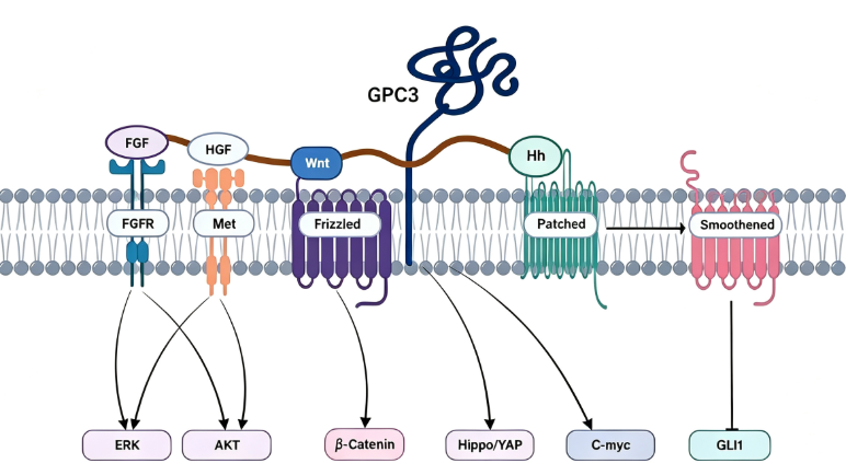
II. Molecular Mechanisms of GPC3 in Hepatocellular Carcinoma Development
GPC3 plays a significant role in cell growth and differentiation by regulating the Wnt signaling pathway, Hedgehog signaling pathway, fibroblast growth factor-2 (FGF-2), and bone morphogenetic protein (BMP) activity. In HCC, GPC3 promotes tumor cell proliferation and survival by modulating the activity of the canonical Wnt/β-catenin signaling pathway. The heparan sulfate side chains of GPC3 synergize with the HGF/c-Met signaling pathway to facilitate HCC cell migration. The HS20 antibody, which targets the HS side chains, inhibits c-Met activation when treating HCC cells, thereby suppressing HGF-mediated migration and metastasis. Knockout of GPC3 significantly reduces the migration and motility of HCC cells, confirming its critical regulatory role in tumor metastasis. GPC3 is released into peripheral blood in a soluble form, and its combination with AFP can further improve the sensitivity of non-invasive diagnosis for liver tumors, making it a dual target for HCC diagnosis and treatment.
III. Clinical Progress in GPC3-Targeted Immunotherapy
GPC3 has emerged as one of the most promising targets for immunotherapy in liver cancer, with multiple therapeutic strategies under clinical development. In the monoclonal antibody field, codrituzumab is the first antibody targeting GPC3. Although its Phase II clinical study did not meet the primary endpoint in the overall population, subgroup analysis showed that patients with high GPC3 expression could benefit from the treatment. In the bispecific antibody field, ERY974 is a CD3/GPC3-targeting bispecific antibody that recruits T cells to tumor sites to exert cytotoxic effects. CM350, developed by Connect Biopharma, is the first GPC3×CD3 bispecific antibody approved for clinical trials in China. In the CAR-T cell therapy field, CARsgen Therapeutics has three GPC3-targeting CAR-T products: CT011 has completed Phase I trials; CT0180 employs antibody-fused TCR technology to reduce cytokine release; and CT0181 co-expresses IL-7 to enhance T cell persistence. ECT204, developed by Eureka Therapeutics, has received FDA orphan drug designation. JWATM204, developed by JW Therapeutics, utilizes the ARTEMIS® platform and has initiated Phase I clinical trials.
IV. Conclusion
As an oncofetal antigen specifically highly expressed in hepatocellular carcinoma, GPC3 has become a core target in HCC diagnosis, imaging, and immunotherapy due to its molecular mechanisms involving the regulation of Wnt, FGF-2, and HGF/c-Met signaling pathways, which promote tumor proliferation, migration, and metastasis. Although no GPC3-targeting drug has been approved globally, diverse approaches, including monoclonal antibodies, bispecific antibodies, and CAR-T cell therapies, are advancing steadily. Alexa Fluor 647-labeled GPC3 recombinant protein provides a reliable detection tool to support related basic research and drug development.
In the development of GPC3-targeted therapies and related research, high-quality fluorescent-labeled recombinant proteins are essential tools for flow cytometry detection and target validation. To meet this research demand, U-Protein offers Alexa Fluor 647-Labeled GPC3 His Tag Protein, Human, suitable for applications such as GPC3 CAR-T cell positivity rate detection, binding activity evaluation of GPC3-targeting antibody drugs, and flow cytometry analysis of GPC3 expression levels on the surface of HCC cell lines.
This article is reviewed and published by the technical expert team of UA
Disclaimer: This article partially utilizes artificial intelligence assistance in its creation. If any content involves copyright or intellectual property issues, please let us know and we promise to verify and remove it as soon as possible.
Purchase recombinant protein, choose Nanjing UA-Bio
UA protein focuses on providing various protein reagents, raw materials, and services required for drug research and development, cell therapy, gene therapy, and basic scientific research, including drug target proteins, immune checkpoint proteins, cytokines, tool enzymes, customized protein expression, and full-length transmembrane protein development. Youai is committed to providing customers with high-quality products and professional services, and building a High-tech Biological Enterprise with International Competitiveness.
Target proteins | membrane proteins | cytokines | enzymes | viral antigens | protein customization
Nanjing UA Biotechnology Co., Ltd. Email:order@ua-bio.com Phone:+86-25-56221161

Product Information

Link:





©2021-2026 all rights reserved UA Biotechnology Co.,Ltd.
Privacy Policy 
网站备案号 浙ICP备2022019033号-2 浙公网安备33010202004867号
 Business license (three certificates in one)
 Tel:+86-25-56221161
E-mail:order@ua-bio.com
All products sold on this website shall not be used for clinical diagnosis or treatment of humans or animals, and shall only be used for non-medical purposes such as industrial or scientific research.
