




Osteoclast differentiation and functional regulation: from physiological bone remodeling to pathological therapeutic targets
Author: Senior Product Manager of UA Views:4 


This article systematically elucidates the physiological functions of osteoclasts in bone remodeling through the secretion of acids and hydrolytic enzymes for bone resorption, focusing on their molecular characteristics and functional regulatory mechanisms. It provides an in-depth analysis of the pivotal regulatory roles of the RANKL/RANK signaling axis and various cytokines (such as M-CSF and IL-6) in osteoclast differentiation, and explores their pathological significance in bone-destructive diseases like osteoporosis.
- Recent Advances
- Product Information
Recent Advances
Osteoclast Differentiation and Functional Regulation: From Physiological Bone Remodeling to Pathological Therapeutic Targets
Summary
This article systematically elaborates on the molecular characteristics and functional regulation mechanisms of osteoclasts, focusing on their physiological role in bone remodeling through the secretion of acids and hydrolytic enzymes for bone resorption. It provides an in-depth analysis of the core regulatory role of the RANKL/RANK signaling axis and various cytokines (M-CSF, IL-6, etc.) in osteoclast differentiation and discusses their pathological significance in bone-destructive diseases such as osteoporosis.
This article systematically elaborates on the molecular characteristics and functional regulation mechanisms of osteoclasts, focusing on their physiological role in bone remodeling through the secretion of acids and hydrolytic enzymes for bone resorption. It provides an in-depth analysis of the core regulatory role of the RANKL/RANK signaling axis and various cytokines (M-CSF, IL-6, etc.) in osteoclast differentiation and discusses their pathological significance in bone-destructive diseases such as osteoporosis.
I. Biological Characteristics and Structure of Osteoclasts
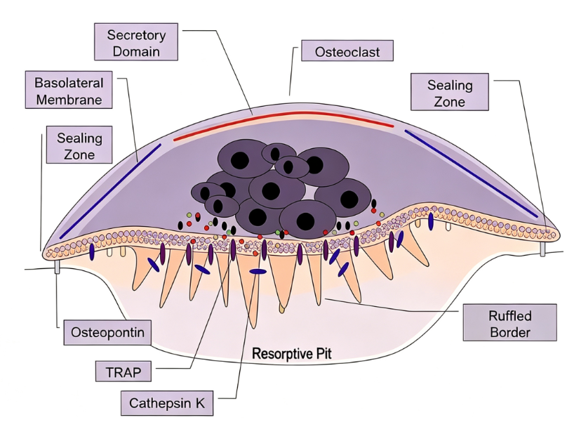
Osteoclasts are large multinucleated cells derived from the self-fusion of monocyte/macrophage lineage cells, exclusively performing bone resorption on bone tissue surfaces. These cells were first discovered by Kolliker in 1873 and are located in concave areas on bone surfaces known as Howship's lacunae. Human osteoclasts typically contain 5 to 20 nuclei, with diameters reaching 150 to 200 μm, and can form even larger cells with diameters up to 100 μm under in vitro induction conditions. Their cytoplasm exhibits a uniform "foamy" appearance due to high concentrations of vesicles and vacuoles, which contain numerous lysosomes filled with acid phosphatase. In active bone resorption states, osteoclasts form a characteristic "ruffled border" membrane structure, which significantly increases cell surface area to facilitate ion secretion and the absorption of matrix degradation products, serving as a morphological marker of actively resorbing osteoclasts.
II. Core Regulatory Signals in Osteoclastogenesis
The differentiation, maturation, and activation of osteoclasts are precisely regulated by various signaling molecules. The RANKL-RANK signaling axis is the most central regulatory pathway in osteoclastogenesis. RANKL (Receptor Activator of Nuclear Factor κB Ligand), a member of the tumor necrosis factor superfamily, is primarily expressed in membrane-bound form by osteoblasts and bone marrow stromal cells, with its expression upregulated under the influence of bone-resorbing stimulants such as 1,25-dihydroxyvitamin D3 and parathyroid hormone. Upon binding to the RANK receptor on osteoclast precursors, RANKL recruits TNF receptor-associated factors, activating key transcription factors such as NF-κB and NFATc1, initiating the transcriptional program of osteoclast-specific genes, and driving the fusion of precursor cells into mature multinucleated osteoclasts.
M-CSF (Macrophage Colony-Stimulating Factor) is another indispensable factor in osteoclastogenesis. Produced by osteoblasts and stromal cells, M-CSF binds to the c-fms receptor on osteoclast precursors, activating downstream signaling pathways to promote the survival and proliferation of osteoclast precursors and upregulating RANK expression to enhance the sensitivity of precursor cells to RANKL. M-CSF gene-deficient op/op mice exhibit an osteopetrosis phenotype, demonstrating its irreplaceable role in osteoclastogenesis. Osteoprotegerin (OPG), a natural decoy receptor for RANKL, is produced and secreted by osteoblasts. It binds to RANKL with high affinity, competitively blocking the interaction between RANKL and RANK, thereby inhibiting osteoclast differentiation and activation, serving as a negative regulator of bone resorption.
III. Molecular Mechanisms of Bone Resorption by Osteoclasts
The bone resorption function of osteoclasts relies on the synergistic effects of an acidic environment and proteolytic enzymes. Osteoclasts generate carbonic acid through the catalysis of carbon dioxide and water by carbonic anhydrase II, which dissociates into hydrogen ions and bicarbonate. These hydrogen ions are actively pumped into the sealed resorption lacuna by highly concentrated vacuolar ATPase proton pumps on the ruffled border membrane, reducing the lacuna pH to approximately 4.5, thereby dissolving mineral components such as hydroxyapatite in bone tissue. Simultaneously, various proteolytic enzymes are released into the resorption lacuna to digest the organic components of the bone matrix. Cathepsin K is the most abundant and functionally important collagen-degrading enzyme in osteoclasts. The 37 kDa proenzyme undergoes autocatalytic cleavage to form the 27 kDa mature active form, which is transported to the ruffled border via intercellular vesicles and secreted into the resorption lacuna to efficiently degrade type I collagen. Inactivating mutations in the cathepsin K gene can lead to pycnodysostosis, further confirming its critical role in bone resorption. MMP-9, a gelatinase essential for osteoclast migration, and MMP-13 also participate in the bone resorption and osteoclast differentiation processes. The degradation products of bone resorption are transported across the cell from the ruffled border to the basolateral membrane via transcytosis and released into circulation, completing the bone resorption process.
IV. Clinical Significance of Osteoclasts and In Vitro Differentiation Models
Abnormally enhanced osteoclast activity is a common pathological basis for bone-destructive diseases such as osteoporosis, Paget's disease, bone metastasis of tumors, and rheumatoid arthritis, as well as a core mechanism of aseptic loosening of artificial joint prostheses. In vitro, the Raw264.7 macrophage cell line can be directionally differentiated into TRAP-positive multinucleated osteoclasts under the synergistic induction of RANKL and M-CSF, providing an important tool for studying osteoclast differentiation mechanisms and screening anti-resorptive drugs. A typical induction protocol involves M-CSF at 40 ng/mL combined with RANKL at 100 ng/mL, with fresh cytokines replenished every 3 days. After 10 days of induction, fused TRAP-positive multinucleated cells can be observed. This in vitro differentiation model has been widely used in osteoclast biology research and anti-osteoporosis drug discovery. To meet this research demand, Uni offers a Raw264.7 osteoclast differentiation cytokine kit, which includes recombinant M-CSF and recombinant RANKL, two highly active cytokines for the directional in vitro induction of Raw264.7 cells into osteoclasts. This kit is suitable for applications such as osteoclast differentiation mechanism research, RANKL/RANK signaling pathway analysis, and in vitro activity evaluation of anti-osteoporosis drugs.
V. Conclusion
As the only cells in the body capable of bone resorption, osteoclasts play an irreplaceable role in bone remodeling and calcium-phosphorus metabolism. Their differentiation is precisely regulated by the RANKL/RANK/OPG signaling axis and synergistically influenced by various cytokines such as M-CSF and IL-6. The in vitro Raw264.7 differentiation model provides an important tool for osteoclast biology research, while the cytokine kit offers a standardized induction protocol for this model.
This article is reviewed and published by the technical expert team of UA
Disclaimer: This article partially utilizes artificial intelligence assistance in its creation. If any content involves copyright or intellectual property issues, please let us know and we promise to verify and remove it as soon as possible.
Purchase recombinant protein, choose Nanjing UA-Bio
UA protein focuses on providing various protein reagents, raw materials, and services required for drug research and development, cell therapy, gene therapy, and basic scientific research, including drug target proteins, immune checkpoint proteins, cytokines, tool enzymes, customized protein expression, and full-length transmembrane protein development. Youai is committed to providing customers with high-quality products and professional services, and building a High-tech Biological Enterprise with International Competitiveness.
Target proteins | membrane proteins | cytokines | enzymes | viral antigens | protein customization
Nanjing UA Biotechnology Co., Ltd. Email:order@ua-bio.com Phone:+86-25-56221161

Product Information

Link:





©2021-2026 all rights reserved UA Biotechnology Co.,Ltd.
Privacy Policy 
网站备案号 浙ICP备2022019033号-2 浙公网安备33010202004867号
 Business license (three certificates in one)
 Tel:+86-25-56221161
E-mail:order@ua-bio.com
All products sold on this website shall not be used for clinical diagnosis or treatment of humans or animals, and shall only be used for non-medical purposes such as industrial or scientific research.
