




PSMA: The Core Target and Molecular Basis for Precision Diagnosis and Treatment of Prostate Cancer
Author: Senior Product Manager of UA Views:4 


This article focuses on the molecular characteristics and biological functions of prostate-specific membrane antigen (PSMA), systematically elaborating its structural features as a type II transmembrane glycoprotein and its specific high expression pattern in prostate cancer cells, while analyzing its core value in the diagnosis, staging, efficacy evaluation, and targeted therapy of prostate cancer.
- Recent Advances
- Product Information
Recent Advances
PSMA: The Core Target and Molecular Basis for Precision Diagnosis and Treatment of Prostate Cancer
Overview
This article systematically elaborates on the molecular characteristics and biological functions of prostate-specific membrane antigen (PSMA), focusing on its structural features as a type II transmembrane glycoprotein and its specific high expression pattern in prostate cancer cells. It analyzes its core value in the diagnosis, staging, efficacy evaluation, and targeted therapy of prostate cancer.
This article systematically elaborates on the molecular characteristics and biological functions of prostate-specific membrane antigen (PSMA), focusing on its structural features as a type II transmembrane glycoprotein and its specific high expression pattern in prostate cancer cells. It analyzes its core value in the diagnosis, staging, efficacy evaluation, and targeted therapy of prostate cancer.
1. Discovery and Molecular Structural Characteristics of PSMA
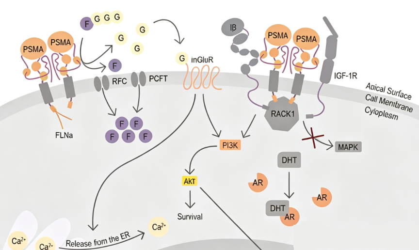
Prostate-specific membrane antigen was first discovered in 1987 as a unique type II transmembrane glycoprotein of prostate epithelial cells and a member of the metallopeptidase family. From a structural biology perspective, PSMA's protein structure consists of three functionally distinct parts: the N-terminal intracellular domain, containing 19 amino acid residues, participates in intracellular signal transduction; the transmembrane domain, composed of 24 amino acid residues, anchors the protein to the lipid bilayer of the cell membrane; and the extracellular domain, containing 707 amino acid residues, is the core region for ligand binding and endocytosis. The extracellular domain of PSMA exhibits metallopeptidase activity, catalyzing the hydrolysis of N-acetyl-aspartyl-glutamate. In normal prostate cells, PSMA is primarily localized in the cytoplasm, whereas in prostate cancer cells, it is abundantly transferred to the cell membrane surface. This unique expression pattern forms the molecular basis for tumor targeting. PSMA exists in various isoforms, including PSM, PSM-B, and PSM-C, all of which are found in the cytoplasm, and their functional significance is still under investigation.
2. Expression Profile and Tumor Specificity of PSMA
PSMA expression exhibits significant tumor-selective characteristics, which are key to its core value in the diagnosis and treatment of prostate cancer. In normal tissues, PSMA is expressed at certain levels in benign prostate epithelial cells, benign prostatic hyperplasia tissues, and prostatic intraepithelial neoplasia, but the expression intensity is far lower than in prostate cancer cells. PSMA is most highly expressed in metastatic and androgen-independent prostate cancer cells, reaching levels 100 to 1000 times higher than in normal cells. PSMA is also overexpressed in newly formed blood vessels of prostate cancer and various solid tumors but is absent in normal tissue neovasculature. Low levels of PSMA expression can also be detected in other normal tissues, including the stomach, bladder, liver, esophagus, colon, breast, fallopian tubes, kidneys, small intestine, lungs, and testes, but these levels are significantly lower than in prostate cancer tissues.
3. Clinical Significance of PSMA in Prostate Cancer
The expression level of PSMA in prostate cancer is closely related to tumor malignancy and clinical progression. Studies have shown that PSMA expression in cancer cells increases with tumor grade and is further enhanced in advanced-stage cancer and anti-androgen-treated cancer cells. Increased PSMA expression is significantly associated with tumor aggressiveness, metastatic potential, and disease recurrence risk, making it an ideal target for ligand-receptor-based imaging and therapy. In diagnostics, PSMA positron emission tomography (PET) has been widely used for the localization of primary lesions, lymph node metastases, and distant metastases in prostate cancer, with superior sensitivity and specificity compared to traditional imaging methods. In therapeutics, 177Lu-PSMA-617 radioligand therapy has been approved for the treatment of metastatic castration-resistant prostate cancer, demonstrating the clinical value of PSMA as a therapeutic target.
4. Biological Properties of PSMA and Implications for Targeting Strategies
The molecular structure of PSMA provides multiple advantages as a target. The extracellular domain of PSMA can link ligands with various functions (e.g., small-molecule inhibitors, antibodies, peptides), while the intracellular domain contains functional factors for endocytosis, initiating intracellular endocytosis and biochemical cycling. This increases the accumulation of radiotracers or drugs inside cells, improving imaging contrast or therapeutic efficacy. The metallopeptidase activity of PSMA also offers possibilities for developing enzyme activity-based molecular probes.
5. Conclusion
PSMA, with its 100- to 1000-fold specific overexpression in prostate cancer cells, increasing expression levels with tumor progression, and endocytic properties following ligand binding, has become the most widely used and successful molecular target in the precision diagnosis and treatment of prostate cancer. From PSMA PET/CT imaging to 177Lu-PSMA radioligand therapy, PSMA-targeted strategies have been integrated into the entire process of prostate cancer diagnosis, staging, treatment, and efficacy evaluation. Human PSMA recombinant protein, as a critical tool for basic research and diagnostic reagent development, will continue to provide essential support for optimizing precision diagnosis and treatment strategies and developing novel targeted drugs for prostate cancer.
In PSMA-related basic research and reagent development, high-quality human PSMA recombinant protein is a key tool for antibody screening, ligand binding analysis, and diagnostic reagent quality control. To meet this research demand, U-Trust offers PSMA/FOLH1 His Tag Protein, Human, suitable for in vitro screening and activity evaluation of anti-PSMA antibody drugs, PSMA ligand binding analysis, and the establishment and validation of PSMA immunoassay methods.
This article is reviewed and published by the technical expert team of UA
Disclaimer: This article partially utilizes artificial intelligence assistance in its creation. If any content involves copyright or intellectual property issues, please let us know and we promise to verify and remove it as soon as possible.
Purchase recombinant protein, choose Nanjing UA-Bio
UA protein focuses on providing various protein reagents, raw materials, and services required for drug research and development, cell therapy, gene therapy, and basic scientific research, including drug target proteins, immune checkpoint proteins, cytokines, tool enzymes, customized protein expression, and full-length transmembrane protein development. Youai is committed to providing customers with high-quality products and professional services, and building a High-tech Biological Enterprise with International Competitiveness.
Target proteins | membrane proteins | cytokines | enzymes | viral antigens | protein customization
Nanjing UA Biotechnology Co., Ltd. Email:order@ua-bio.com Phone:+86-25-56221161

Product Information

Link:





©2021-2026 all rights reserved UA Biotechnology Co.,Ltd.
Privacy Policy 
网站备案号 浙ICP备2022019033号-2 浙公网安备33010202004867号
 Business license (three certificates in one)
 Tel:+86-25-56221161
E-mail:order@ua-bio.com
All products sold on this website shall not be used for clinical diagnosis or treatment of humans or animals, and shall only be used for non-medical purposes such as industrial or scientific research.
