




TNFR-1/CD120a: Key Receptor in TNF Signaling Pathway and Core Regulator of Inflammation
Author: Senior Product Manager of UA Views:23 


This article focuses on the molecular structure and biological functions of TNFR-1 (CD120a), systematically elaborating its classification as a core member of the tumor necrosis factor receptor superfamily, structural characteristics, and its key roles in inflammatory signal transduction and apoptosis regulation.
- Recent Advances
- Product Information
Recent Advances
TNFR-1/CD120a: The Key Receptor in TNF Signaling Pathway and Core Regulator of Inflammation
Overview
This article systematically elaborates on the molecular structure and biological functions of TNFR-1 (CD120a), detailing its classification as a core member of the tumor necrosis factor receptor superfamily, structural characteristics, and its pivotal role in inflammatory signal transduction and apoptosis regulation.
This article systematically elaborates on the molecular structure and biological functions of TNFR-1 (CD120a), detailing its classification as a core member of the tumor necrosis factor receptor superfamily, structural characteristics, and its pivotal role in inflammatory signal transduction and apoptosis regulation.
I. Molecular Classification and Family Positioning of TNFR-1
Tumor necrosis factor receptor-1 (TNFR-1), also known as CD120a or TNFRSF1A, is a key member of the nerve growth factor receptor (NGFR) superfamily. This superfamily includes NGFR, TNFR-1 (CD120a), TNFR-II (CD120b), CD40, CD27, T-cell cDNA-41BB encoded product, rat T-cell antigen OX40, and human myeloid cell surface activation antigen Fas (CD95). TNFR-1 and TNFR-II belong to the TNF receptor family, sharing homology in their extracellular domains but exhibiting significant differences in intracellular signal transduction. TNFR-1 contains a death domain (DD) in its intracellular region, enabling it to mediate apoptotic signals, whereas TNFR-II lacks this structure.
II. Structural Features and Ligand Binding Mechanism of TNFR-1
TNFR-1 is a type I transmembrane glycoprotein with an extracellular region composed of four cysteine-rich domains, each containing approximately 40 amino acid residues with 4 to 6 conserved cysteine residues. These cysteines form disulfide bonds to stabilize the receptor's three-dimensional conformation, serving as the structural basis for ligand binding. The first N-terminal region of TNFR-1 contains six conserved cysteines and one each of Tyr, Gly, and Thr residues, while other regions also contain 4 to 6 cysteines. TNFR-1 exhibits ligand binding specificity for both TNF-α and TNF-β (lymphotoxin-α), with TNF-α being its primary physiological ligand. TNF-α binds to TNFR-1 as a homotrimer, inducing receptor trimerization, which is a critical step in initiating downstream signal transduction. The affinity between TNF-α and TNFR-1 is in the nanomolar range, ensuring the adjustability of signal transmission.
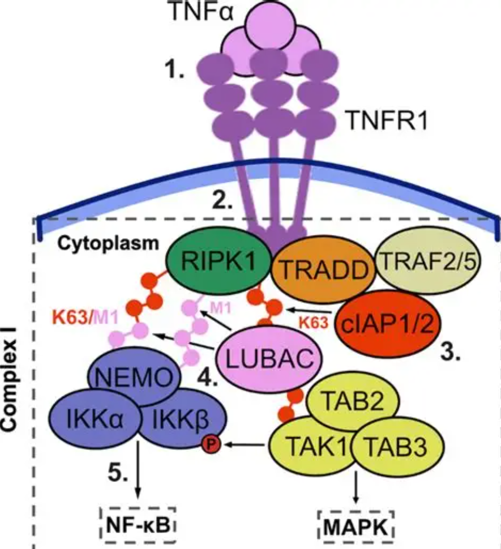
III. Signal Transduction Pathways Mediated by TNFR-1
The intracellular region of TNFR-1 contains an approximately 80-amino acid death domain, which is the core functional element distinguishing TNFR-1 from TNFR-II. Upon TNF-α binding, TNFR-1 undergoes trimerization, leading to the aggregation of the death domain and subsequent recruitment of the adaptor protein TRADD (TNFR1-associated death domain protein). TRADD acts as a platform molecule, further recruiting various signaling molecules to form two functionally distinct signal complexes. Complex I forms near the cell membrane and includes RIPK1, TRAF2, and cIAPs, primarily activating the NF-κB and MAPK pathways to promote the expression of pro-inflammatory cytokines and cell survival genes, mediating inflammatory responses. Upon dissociation of Complex I, TRADD forms Complex II (death-inducing signaling complex) with FADD and pro-caspase-8, activating the caspase cascade and inducing apoptosis. The signaling pathways mediated by TNFR-1 play a central role in inflammation regulation, immune responses, and cellular homeostasis maintenance. Dysregulation of these pathways is closely associated with rheumatoid arthritis, inflammatory bowel disease, and various autoimmune disorders. Targeted therapeutic strategies based on TNFR-1 signaling have been extensively studied and applied in various inflammatory diseases.
IV. Soluble Form of TNFR-1 and Its Pathological Significance
TNFR-1 exists in a soluble form (sTNFR-1), generated through proteolytic cleavage of the membrane-bound receptor, and can be detected in serum and body fluids. sTNFR-1 competitively binds TNF-α, acting as a natural antagonist. Elevated levels of sTNFR-1 are observed in various inflammatory and autoimmune diseases and are closely correlated with disease activity. The detection of sTNFR-1 has shown potential value in assessing conditions such as rheumatoid arthritis, sepsis, and heart failure.
V. Conclusion
As a key receptor in the TNF signaling pathway, TNFR-1 plays a central role in immune regulation and inflammatory responses through its dual functions in mediating inflammation and apoptosis. Its structural features—the extracellular cysteine-rich domains and intracellular death domain—determine its unique ligand recognition and signal transduction properties. Human recombinant TNFR-1 protein, as an essential tool for basic research and drug development, will continue to provide critical support for in-depth analysis of TNFR-1 signaling networks and the exploration of related disease treatment strategies.
In TNFR-1-related basic research and drug screening, high-quality human recombinant TNFR-1 protein is a core tool for ligand binding analysis, signal pathway studies, and antagonist screening. To meet this research demand, Uni provides TNFR-1/CD120a Protein, Human, suitable for applications such as TNF-α and TNFR-1 binding activity analysis, TNFR-1 signal transduction mechanism research, and anti-TNFR-1 antibody drug activity evaluation.
This article is reviewed and published by the technical expert team of UA
Disclaimer: This article partially utilizes artificial intelligence assistance in its creation. If any content involves copyright or intellectual property issues, please let us know and we promise to verify and remove it as soon as possible.
Purchase recombinant protein, choose Nanjing UA-Bio
UA protein focuses on providing various protein reagents, raw materials, and services required for drug research and development, cell therapy, gene therapy, and basic scientific research, including drug target proteins, immune checkpoint proteins, cytokines, tool enzymes, customized protein expression, and full-length transmembrane protein development. Youai is committed to providing customers with high-quality products and professional services, and building a High-tech Biological Enterprise with International Competitiveness.
Target proteins | membrane proteins | cytokines | enzymes | viral antigens | protein customization
Nanjing UA Biotechnology Co., Ltd. Email:order@ua-bio.com Phone:+86-25-56221161

Product Information

Link:





©2021-2026 all rights reserved UA Biotechnology Co.,Ltd.
Privacy Policy 
网站备案号 浙ICP备2022019033号-2 浙公网安备33010202004867号
 Business license (three certificates in one)
 Tel:+86-25-56221161
E-mail:order@ua-bio.com
All products sold on this website shall not be used for clinical diagnosis or treatment of humans or animals, and shall only be used for non-medical purposes such as industrial or scientific research.
