




Classification, phenotypic markers, and functional immune characteristics of dendritic cell subsets
Author: Senior Product Manager of UA Views:13 


This article systematically elaborates on the subset-specific surface markers, TLR expression profiles, and cytokine secretion characteristics of human and mouse-derived conventional DCs (cDC1 and cDC2), plasmacytoid DCs, and monocyte-derived DCs, focusing on the classification system of dendritic cells.
- Recent Advances
- Product Information
Recent Advances
Classification, Phenotypic Markers, and Functional Characteristics of Dendritic Cell Subsets
Summary
This article systematically elaborates on the subset-specific surface markers, TLR expression profiles, and cytokine secretion characteristics of human and murine conventional DCs (cDC1 and cDC2), plasmacytoid DCs, and monocyte-derived DCs.
This article systematically elaborates on the subset-specific surface markers, TLR expression profiles, and cytokine secretion characteristics of human and murine conventional DCs (cDC1 and cDC2), plasmacytoid DCs, and monocyte-derived DCs.
I. General Framework of DC Subset Classification
As the most potent antigen-presenting cells in the body, dendritic cells can be divided into multiple subsets based on their developmental origin, phenotypic characteristics, and functional differences. cDC1 and cDC2 are conventional DCs, forming the core population for immune surveillance and antigen presentation. pDCs are characterized by their secretion of type I and III interferons and play a unique role in antiviral immunity. MoDCs primarily exist under inflammatory conditions, differentiating from monocytes, and exhibit high plasticity.
II. Phenotypic Markers and Functions of Human cDC1
Human cDC1 is characterized by the expression of CD141⁺CD11c⁺ (signature combination), along with high levels of HLA-DR, XCR1, CLEC9A, DEC205, and CADM1, and low levels of CD11c. Their TLR expression profile includes TLR1, 3, 6, 8, and 10, along with STING and CLEC12A. The core function of cDC1 is to activate CD8⁺ T cells and Th1 immune responses through cross-presentation, correlating with favorable prognosis in antitumor immunity. Their secretion profile includes high levels of IL-12, TNF-α, CXCL9, and CXCL10.
III. Phenotypic Markers and Functions of Murine cDC1
Murine cDC1 is marked by the signature combination of CD11c⁺CD24⁺, along with MHC-II, XCR1, CLEC9A, and DEC205. CD8α⁺ (resident) and CD103⁺ (migratory) are its main subset markers. The TLR expression profile includes TLR2⁺, TLR4, TLR11⁺, TLR13, and STING. Functionally, murine cDC1 also primarily activates CD8⁺ T cells and Th1 immune responses through cross-presentation, but it exhibits stronger secretion capabilities for IL-12, type I, and type III IFNs.
IV. Phenotypic Markers and Functions of Human cDC2
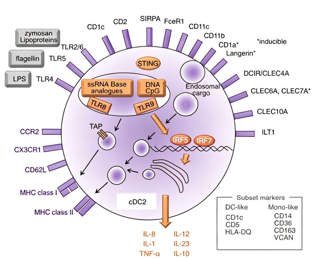
Human cDC2 is characterized by the signature combination of CD1c⁺CD11c⁺, along with HLA-DR, CD11b, CD172a (SIRPα), CD1a (migratory), and subset markers CD14 and CD5. cDC2 exhibits diverse functions, inducing the activation of Th17, Th1, Th2, Treg, and CD8⁺ T cells, and plays a role in maintaining Treg-Th17 homeostasis in the gut and lungs. Its secretion profile includes various pro- and anti-inflammatory cytokines such as IL-12, IL-1, IL-8, TNF-α, IL-10, and IL-23.
V. Phenotypic Markers and Functions of pDC
Human pDC is marked by the signature combination of CD123⁺CD303⁺ (CLEC4C)CD304⁺, along with CCR2 and CXCR3, and low levels of CD11c and HLA-DR. TLR7 and TLR9 are highly expressed, along with RLR, STING, and CLEC12A. During acute or chronic viral infections, pDCs primarily secrete type I and III interferons and can activate CD8⁺ T cells through cross-presentation. In tumor immunity, pDCs are typically associated with poor prognosis and contribute to disease progression in autoimmune disorders. Their secretion profile is dominated by IFN-α, along with IL-6, granzyme B, and TNF-α. Murine pDCs are characterized by B220⁺CD317⁺SIGLEC-H⁺, low CD11c and MHC-II, and high TLR7 and TLR9 expression.
VI. Phenotypic Markers and Functions of MoDC
Human MoDC is characterized by the signature combination of CD11c⁺HLA-DR⁺CD1c⁺CD11b⁺, along with CD14, CD64, CD206, CD209, CD172a, and CCR2. Murine MoDC is marked by CD11c⁺MHC-II⁺CD11b⁺Ly6C⁺, along with CD64, CD206, CD209, and CD14. MoDCs primarily differentiate from monocytes during inflammatory responses, promoting the differentiation of CD4⁺ T cells into Th1, Th2, or Th17 subsets, exhibiting high plasticity.
VII. Conclusion
As a critical bridge between innate and adaptive immunity, the functional diversity of dendritic cells relies on the coordinated roles of subsets such as cDC1, cDC2, pDC, and MoDC. Each subset executes distinct immune functions through characteristic surface markers, TLR expression profiles, and cytokine secretion patterns. A comprehensive understanding of DC subset classification and functional characteristics is essential for DC-related immunological research and the design of precise immune intervention strategies.
In DC subset differentiation and functional studies, standardized in vitro induction systems are key to obtaining specific DC subsets. To meet this research need, UniLove provides LYPD3/C4.4A His Tag Protein, Mouse, suitable for the in vitro directional induction and differentiation of murine bone marrow-derived dendritic cells. It can be used for DC subset development mechanism studies, antigen presentation function analysis, and immune regulation evaluation.
This article is reviewed and published by the technical expert team of UA
Disclaimer: This article partially utilizes artificial intelligence assistance in its creation. If any content involves copyright or intellectual property issues, please let us know and we promise to verify and remove it as soon as possible.
Purchase recombinant protein, choose Nanjing UA-Bio
UA protein focuses on providing various protein reagents, raw materials, and services required for drug research and development, cell therapy, gene therapy, and basic scientific research, including drug target proteins, immune checkpoint proteins, cytokines, tool enzymes, customized protein expression, and full-length transmembrane protein development. Youai is committed to providing customers with high-quality products and professional services, and building a High-tech Biological Enterprise with International Competitiveness.
Target proteins | membrane proteins | cytokines | enzymes | viral antigens | protein customization
Nanjing UA Biotechnology Co., Ltd. Email:order@ua-bio.com Phone:+86-25-56221161

Product Information

Link:





©2021-2026 all rights reserved UA Biotechnology Co.,Ltd.
Privacy Policy 
网站备案号 浙ICP备2022019033号-2 浙公网安备33010202004867号
 Business license (three certificates in one)
 Tel:+86-25-56221161
E-mail:order@ua-bio.com
All products sold on this website shall not be used for clinical diagnosis or treatment of humans or animals, and shall only be used for non-medical purposes such as industrial or scientific research.
