




In vitro induction, differentiation, and culture strategies of dendritic cells
Author: Senior Product Manager of UA Views:9 


This article systematically elaborates on the in vitro culture requirements of dendritic cells, detailing their developmental origins and subgroup characteristics, while analyzing the technical pathways and operational key points for inducing DC differentiation from various sources such as bone marrow CD34+ hematopoietic stem cells, peripheral blood monocytes, and induced pluripotent stem cells.
- Recent Advances
- Product Information
Recent Advances
In Vitro Induction and Differentiation Strategies for Dendritic Cells
Summary
This article systematically elaborates on the developmental origins and subset characteristics of dendritic cells (DCs) in response to in vitro culture requirements. It analyzes technical pathways and operational key points for inducing DC differentiation from various sources, including bone marrow CD34+ hematopoietic stem cells, peripheral blood monocytes, and induced pluripotent stem cells.
This article systematically elaborates on the developmental origins and subset characteristics of dendritic cells (DCs) in response to in vitro culture requirements. It analyzes technical pathways and operational key points for inducing DC differentiation from various sources, including bone marrow CD34+ hematopoietic stem cells, peripheral blood monocytes, and induced pluripotent stem cells.
I. Developmental Origins and Functional Positioning of Dendritic Cells
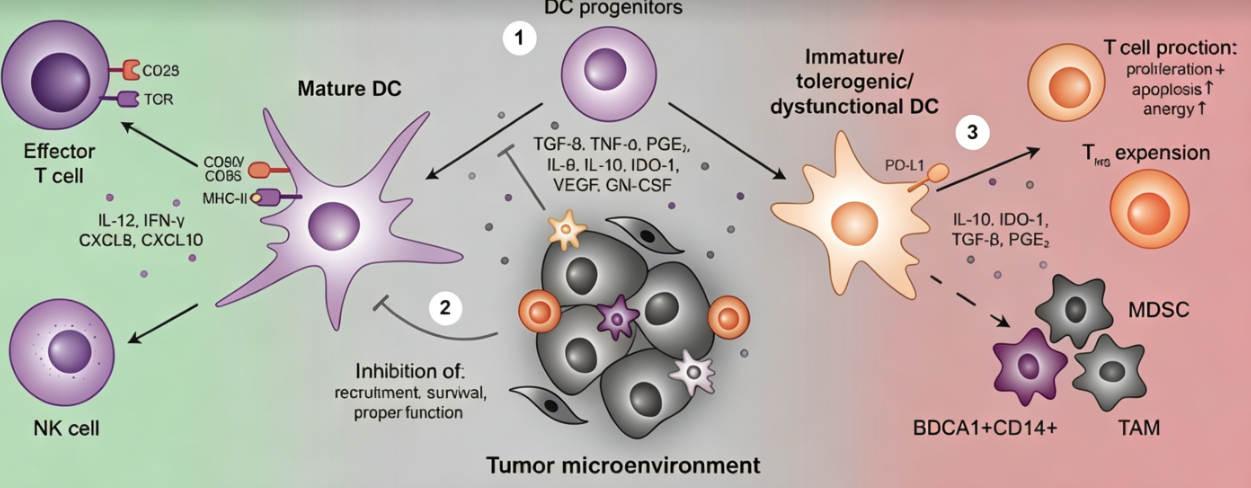
Dendritic cells were first isolated from mouse spleen by researchers in 1973 and are currently recognized as the most potent antigen-presenting cells in the body. A single DC can activate 100 to 3000 T cells, with an antigen-presenting efficiency 100 to 1000 times that of macrophages and B cells. By processing tumor antigens and presenting them to T cells, DCs bridge innate and adaptive immunity, initiating anti-tumor immune responses. Therefore, DCs are regarded as the initiators of immune responses. Most DCs are generated from common DC progenitors under FLT3L induction, ultimately differentiating into classical DCs and plasmacytoid DCs. Some DCs originate from hematopoietic stem cell-derived monocytes, which produce monocyte-derived DCs under the induction of GM-CSF and IL-4. In vitro DC culture sources include bone marrow, peripheral blood monocytes, CD34+ hematopoietic stem cells, or direct isolation from blood and tissue samples. Induced pluripotent stem cells can also be used to obtain DCs.
II. Induction of DC Differentiation from Human Bone Marrow CD34+ Hematopoietic Stem Cells
Inducing DC differentiation from human bone marrow CD34+ hematopoietic stem cells requires pre-seeding OP9 feeder cells (density of 2.5×10⁴ cells/mL, 5000 cells per well in a 96-well plate). CD34+ cells are seeded at a density of 3000 cells per well onto the OP9 feeder layer. Starting from day 6, perform half-medium changes by adding DC differentiation medium containing Flt3L (100 ng/mL), SCF (20 ng/mL), and GM-CSF (20 μg/mL). Cells can be harvested between days 12 and 21, with half-medium changes performed on days 12 and 18. This protocol can induce the generation of multiple DC subsets.
III. Induction of MoDC Differentiation from Human Peripheral Blood Monocytes
Enriched monocytes are plated in a 24-well plate at a density of 5×10⁶ cells/mL in RPMI 1640 medium supplemented with 10% FBS, recombinant human IL-4 (500 U/mL), and GM-CSF (500 U/mL), and cultured at 37°C with 5% CO₂. After 48 hours, perform a half-medium change and replenish IL-4 and GM-CSF at the same concentrations. Continue culturing for another 2 days. Immature MoDCs can be obtained by day 5. To obtain mature DCs, stimulate with LPS (0.5 μg/mL) or TNF-α (10 ng/mL) combined with IL-6 (10 ng/mL), IL-1β (10 ng/mL), and PGE2 (1 μmol/L) for 24 to 48 hours. This is the most widely used and operationally convenient DC induction protocol.
IV. Directed Differentiation of Induced Pluripotent Stem Cells into DCs
Differentiation of iPSCs or ESCs into DCs involves two stages. The first stage is differentiation into hematopoietic progenitor cells. When iPSCs reach 70% to 80% confluency, they are digested with collagenase IV to form cell clusters containing 50 to 100 cells. These are cultured under hypoxic conditions to form embryoid bodies, and differentiation is guided using staged induction media. In the second stage, harvested hematopoietic progenitor cells are seeded onto irradiated OP9 feeder layers at a density of 1×10⁶ to 1.5×10⁶ cells/mL to initiate DC differentiation. Adding FLT3L (100 ng/mL), SCF (20 ng/mL), GM-CSF (20 ng/mL), and IL-4 (20 ng/mL) can generate cDC1 and cDC2 subsets, while adding FLT3L (100 ng/mL), SCF (20 ng/mL), and GM-CSF (10 ng/mL) can generate cDC1 and pDC subsets. Perform half-medium changes every two days, and DC morphology may appear by days 4 to 7 of differentiation.
V. Conclusion
In vitro culture of dendritic cells involves a complete technical chain from sample source selection and induction differentiation protocol determination to subset identification. Depending on experimental needs, bone marrow CD34+ hematopoietic stem cells, peripheral blood monocytes, or iPSC-derived sources can be selected as starting materials. Combined with appropriate cytokine combinations and culture conditions, specific DC subsets can be directionally obtained. Standardized cytokine tools are of great significance for improving the success rate and experimental reproducibility of DC culture.
In the aforementioned in vitro DC induction and differentiation studies, high-quality cytokine combinations are key to ensuring the efficiency of directional differentiation and functional integrity of DCs. To meet this research demand, U-Antibody provides the Dendritic Cell Cytokine Set, Human, suitable for in vitro induction of MoDC differentiation from human peripheral blood monocytes, differentiation of DC subsets from human CD34+ hematopoietic stem cells, and directed differentiation of iPSC/ESC-derived hematopoietic progenitor cells into DCs, among other experimental scenarios.
This article is reviewed and published by the technical expert team of UA
Disclaimer: This article partially utilizes artificial intelligence assistance in its creation. If any content involves copyright or intellectual property issues, please let us know and we promise to verify and remove it as soon as possible.
Purchase recombinant protein, choose Nanjing UA-Bio
UA protein focuses on providing various protein reagents, raw materials, and services required for drug research and development, cell therapy, gene therapy, and basic scientific research, including drug target proteins, immune checkpoint proteins, cytokines, tool enzymes, customized protein expression, and full-length transmembrane protein development. Youai is committed to providing customers with high-quality products and professional services, and building a High-tech Biological Enterprise with International Competitiveness.
Target proteins | membrane proteins | cytokines | enzymes | viral antigens | protein customization
Nanjing UA Biotechnology Co., Ltd. Email:order@ua-bio.com Phone:+86-25-56221161

Product Information

Link:





©2021-2026 all rights reserved UA Biotechnology Co.,Ltd.
Privacy Policy 
网站备案号 浙ICP备2022019033号-2 浙公网安备33010202004867号
 Business license (three certificates in one)
 Tel:+86-25-56221161
E-mail:order@ua-bio.com
All products sold on this website shall not be used for clinical diagnosis or treatment of humans or animals, and shall only be used for non-medical purposes such as industrial or scientific research.
