




Activin A and Cancer Cachexia: Vascular Endothelial Injury as a Novel Perspective on Muscle Atrophy
Author: Senior Product Manager of UA Views:15 


This article systematically elucidates the central role of Activin A in the development and progression of cancer cachexia, focusing on the molecular mechanism by which this cytokine drives skeletal muscle atrophy through inhibiting PGC1α expression in vascular endothelial cells and inducing endothelial dysfunction.
- Recent Advances
- Product Information
Recent Advances
Activin A and Cancer Cachexia: Vascular Endothelial Injury as a Novel Perspective on Muscle Atrophy
Summary
This article focuses on the central role of Activin A in the development of cancer cachexia, systematically elucidating the molecular mechanism by which this cytokine drives skeletal muscle atrophy by inhibiting PGC1α expression in vascular endothelial cells and inducing endothelial dysfunction.
This article focuses on the central role of Activin A in the development of cancer cachexia, systematically elucidating the molecular mechanism by which this cytokine drives skeletal muscle atrophy by inhibiting PGC1α expression in vascular endothelial cells and inducing endothelial dysfunction.
I. Pathological Features and Research Challenges of Cancer Cachexia.
Cancer cachexia is a systemic wasting syndrome characterized by involuntary weight loss, skeletal muscle atrophy, and fat depletion, and is one of the most common complications in advanced cancer patients. The occurrence of cachexia not only severely affects patients' quality of life and physical condition but also reduces their tolerance to chemotherapy and radiotherapy. Approximately 20% to 30% of cancer deaths are directly or indirectly related to cachexia. For a long time, research has focused on the atrophy mechanisms within muscle cells themselves, with the "inflammatory cytokine hypothesis" suggesting that inflammatory factors released by tumors directly act on skeletal muscle to induce catabolism. However, the key process by which these pathogenic factors from tumor sites remotely invade muscle tissue through the bloodstream has long remained unclear. Given that skeletal muscle is a highly vascularized tissue, vascular endothelial cells, as the first point of contact with circulating factors, should logically serve as the critical link in sensing pathogenic signals and mediating muscle atrophy.
II. Discovery of Activin A and Vascular Endothelial Dysfunction.
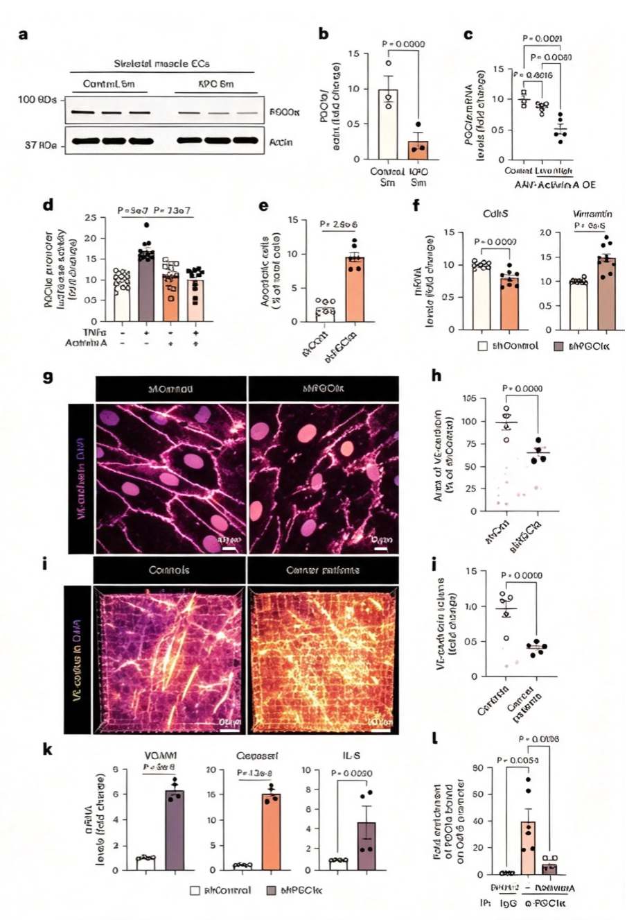
A study published in *Nature Cancer* by researchers at the University of Illinois at Chicago revealed a long-overlooked key link in cancer cachexia. Using KPC transgenic pancreatic cancer mouse models, CT26 colon cancer, LLC1 lung cancer, and B16F10 melanoma models, the team employed tissue-clearing three-dimensional imaging technology to demonstrate that, before any detectable muscle atrophy, there was already a significant reduction in vascular density and vascular fragmentation in mouse skeletal muscle. Clinically, abdominal muscle biopsy samples from cancer patients similarly confirmed reduced vascular density, with more pronounced changes in cachectic patients. These findings established the temporal sequence of "vascular endothelial damage preceding muscle atrophy."
Further transcriptomic analysis of muscle vascular endothelial cells revealed that genes related to muscle cell differentiation were significantly altered in endothelial cells as early as the initial stages of tumor growth, accompanied by changes in the expression of markers for endothelial-mesenchymal transition (EndMT). Through cell clustering analysis, the researchers identified eight distinct endothelial cell subpopulations, several of which were highly sensitive to circulating activin A signals. Quantitative assays confirmed that circulating activin A levels were significantly elevated in multiple cachexia mouse models, and endothelial cells expressed more activin A receptors, suggesting their greater susceptibility to circulating activin A compared to skeletal muscle cells themselves.
III. Molecular Mechanism: The Activin A-PGC1α Axis-Mediated Endothelial Injury.
To validate the direct pathogenic role of activin A, researchers used adeno-associated viruses to overexpress activin A in mice, finding that high-dose activin A significantly reduced muscle vascular density, upregulated pro-apoptotic genes and EndMT markers, and downregulated anti-apoptotic genes and endothelial markers. In early-stage tumor-bearing mice without cachexia symptoms, the number of apoptotic endothelial cells and the proportion of cells undergoing EndMT were already significantly increased, indicating that endothelial injury is an early event in cachexia progression.
The researchers discovered that the expression of PGC1α, a critical regulator of endothelial homeostasis, was significantly downregulated in muscle endothelial cells of cachectic mice. In vivo and in vitro experiments confirmed that activin A directly inhibits PGC1α promoter activity. Mechanistically, under normal conditions, PGC1α binds to the promoter of the Cdh5 gene encoding VE-cadherin to maintain vascular barrier integrity, whereas activin A stimulation blocks this binding, leading to reduced VE-cadherin expression. PGC1α knockdown directly increased endothelial cell apoptosis, induced EndMT, and upregulated multiple pro-inflammatory genes. Using endothelial-specific PGC1α knockout mice, it was demonstrated that even in the absence of tumors, PGC1α downregulation alone could induce cachexia-like phenotypes, including weight loss, muscle atrophy, and vascular leakage.
IV. Conclusion.
Activin A plays a key upstream role in the development of cancer cachexia by inhibiting PGC1α expression in vascular endothelial cells, inducing endothelial cell apoptosis, endothelial-mesenchymal transition, and vascular barrier disruption. The established temporal sequence of vascular endothelial damage preceding skeletal muscle atrophy provides a new window for early intervention in cachexia. Targeting the Activin A-PGC1α axis to restore muscle vascular function may emerge as a novel strategy for treating cancer cachexia.
In studies related to Activin A mechanisms, neutralizing antibody screening, and receptor binding analysis, high-quality human Activin A recombinant protein is a critical experimental tool. To meet this research need, UniLove offers Activin A Protein, Human, suitable for studying Activin A binding activity with its receptors (ActRIIA/ActRIIB), in vitro screening and activity evaluation of anti-Activin A neutralizing antibodies, and exploration of Activin A signaling pathways (Smad2/3 phosphorylation).
This article is reviewed and published by the technical expert team of UA
Disclaimer: This article partially utilizes artificial intelligence assistance in its creation. If any content involves copyright or intellectual property issues, please let us know and we promise to verify and remove it as soon as possible.
Purchase recombinant protein, choose Nanjing UA-Bio
UA protein focuses on providing various protein reagents, raw materials, and services required for drug research and development, cell therapy, gene therapy, and basic scientific research, including drug target proteins, immune checkpoint proteins, cytokines, tool enzymes, customized protein expression, and full-length transmembrane protein development. Youai is committed to providing customers with high-quality products and professional services, and building a High-tech Biological Enterprise with International Competitiveness.
Target proteins | membrane proteins | cytokines | enzymes | viral antigens | protein customization
Nanjing UA Biotechnology Co., Ltd. Email:order@ua-bio.com Phone:+86-25-56221161

Product Information

Link:





©2021-2026 all rights reserved UA Biotechnology Co.,Ltd.
Privacy Policy 
网站备案号 浙ICP备2022019033号-2 浙公网安备33010202004867号
 Business license (three certificates in one)
 Tel:+86-25-56221161
E-mail:order@ua-bio.com
All products sold on this website shall not be used for clinical diagnosis or treatment of humans or animals, and shall only be used for non-medical purposes such as industrial or scientific research.
