

UA-MHC H-2Kb/KSPWFTTL Murine Leukemia Virus p15E Tetramer-APC is a core tool for antigen-specific T cell detection
Views:10 


Immunological significance of the antigenic epitope of murine leukemia virus p15E.
- Recent Advances
- Product Information
Recent Advances
UA-MHC H-2Kb/KSPWFTTL Murine Leukemia Virus p15E Tetramer-APC: A Core Tool for Antigen-Specific T Cell Detection
Immunological Significance of Murine Leukemia Virus p15E Epitope.
Murine leukemia virus (MuLV) is a retrovirus with oncogenic potential in murine hosts, and its research history spans the intersection of tumor virology and immunology. Among the various structural proteins encoded by MuLV, the p15E protein, as the transmembrane subunit of the envelope glycoprotein, plays a key role in viral entry into host cells and is also an important target antigen for the host's antiviral immune response. The nonapeptide sequence KSPWFTTL (also known as the M8 epitope), located at amino acids 604-611 of the p15E protein, has been identified as an immunodominant CD8+ cytotoxic T lymphocyte (CTL) epitope in H-2Kb haplotype mice.
The immunological significance of this epitope extends beyond virology. Studies have shown that the p15E protein can be endogenously expressed in various H-2b haplotype tumor cells, including common tumor models such as B16 melanoma, MC38 colorectal adenocarcinoma, and MCA205 sarcoma cells. Therefore, M8 epitope-specific CD8+ T cells can be effectively detected in tumor-infiltrating lymphocytes, making the KSPWFTTL/H-2Kb system an important bridge connecting antiviral immunity and antitumor immunity research. Notably, although the wild-type p15E epitope has limitations in vaccine immunogenicity, its value as a marker for tumor immune responses has been widely recognized, and optimization studies of related mimotopes continue to advance the field.
Core Principles and Detection Advantages of MHC-Peptide Tetramer Technology.
Accurate quantitative detection and phenotypic analysis of antigen-specific CD8+ T cells are key to understanding adaptive immune response mechanisms. MHC-peptide tetramer technology has been recognized as the gold standard in this field. The core design principle of this technology is based on the specific recognition between the T cell receptor (TCR) and the MHC-peptide complex: biotinylated MHC class I molecules are recombinantly expressed in vitro and correctly folded with specific antigen peptides to form pMHC monomer complexes. The high-affinity binding between streptavidin and biotin is then used to assemble four pMHC monomers into a tetramer structure.
This tetramer structure can simultaneously bind multiple TCRs on the T cell surface, significantly enhancing binding affinity and enabling stable capture of low-affinity T cells. After conjugation with fluorescent dyes, this probe can be used for precise quantitative detection of CD8+ T cells reactive to specific epitopes via flow cytometry. Since this method directly relies on the physical binding of TCR to pMHC complexes and does not involve T cell activation or effector function, it can comprehensively reflect the true frequency of antigen-specific T cell populations, including functionally impaired or resting cells. Whole blood, peripheral blood mononuclear cells, and expanded cell lines are all suitable sample types for tetramer analysis.
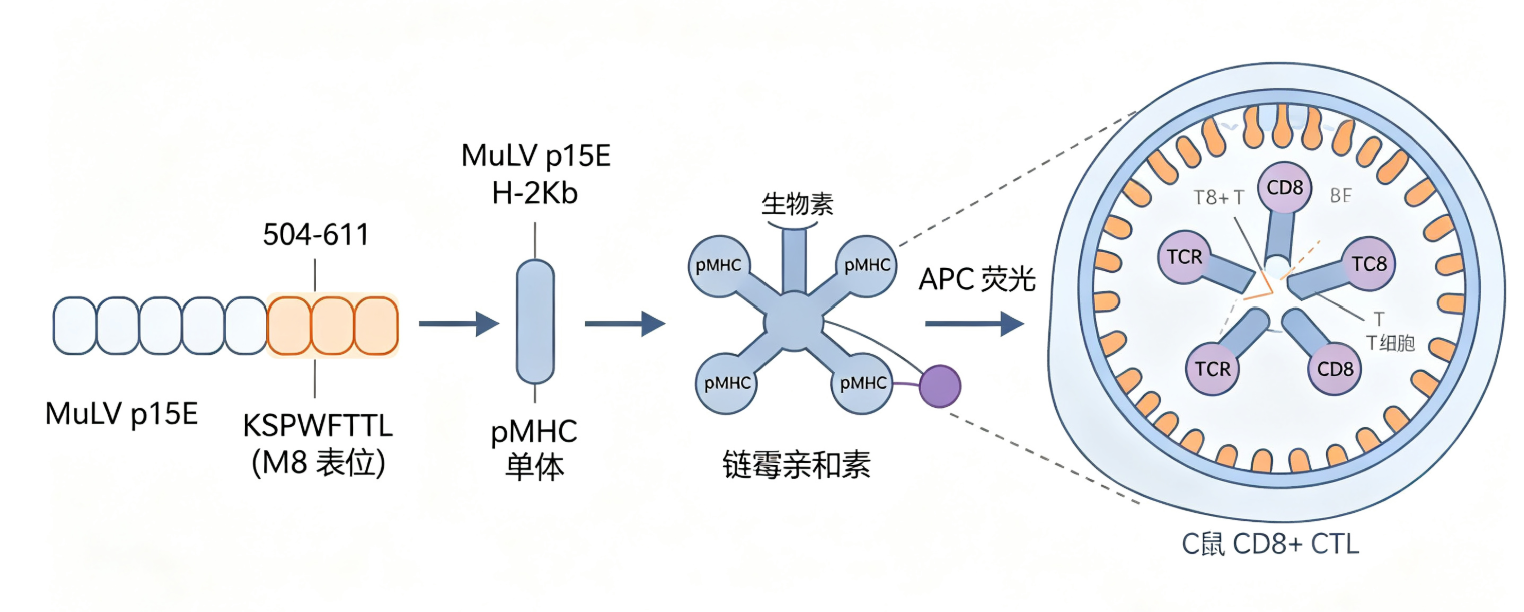
Product Characteristics and Key Technical Parameters of UA-MHC H-2Kb/KSPWFTTL MuLV p15E Tetramer-APC.
To address the above research needs, UA-Biotech offers the high-performance UA-MHC H-2Kb/KSPWFTTL MuLV p15E Tetramer-APC. The design and preparation of this product strictly adhere to the core technical standards of MHC tetramers. In terms of molecular composition, the reagent contains the murine MHC class I molecule H-2Kb allele product, pre-loaded with the KSPWFTTL antigen peptide (corresponding to amino acids 604-611) derived from the MuLV p15E protein. The peptide-H-2Kb complex is assembled into a tetramer, ensuring stability and affinity for specific TCR binding. For fluorescent labeling, the tetramer is conjugated with allophycocyanin (APC), which has high quantum yield and excellent photostability, producing bright and stable fluorescent signals that are highly compatible with standard flow cytometry detection channels, facilitating the design of multicolor fluorescence analysis schemes. The product is supplied in 50-test quantities and should be stored at 4°C protected from light. In terms of applications, this tetramer reagent is suitable for flow cytometric detection of H-2Kb-restricted, MuLV p15E (KSPWFTTL)-specific CD8+ T cells in mouse-derived samples such as peripheral blood, spleen, lymph nodes, and tumor-infiltrating lymphocytes. The product has received positive feedback from users regarding its high specificity and low background staining.
Experimental Application Strategies and Considerations for H-2Kb/KSPWFTTL Tetramer-APC.
The following key strategies should be noted when using the UA-MHC H-2Kb/KSPWFTTL MuLV p15E Tetramer-APC in experiments. First, when selecting anti-mouse CD8 antibodies, caution should be exercised as certain clones (e.g., 53-6.7) may cause high background staining in all CD8+ T cells rather than specifically recognizing antigen-specific T cells. The KT15 clone is recommended for optimal staining results. Second, a checkerboard titration of the tetramer and anti-CD8 antibody is advised to determine the optimal working concentration for specific experimental systems. Typically, controlling cell density within 1×10⁶ to 5×10⁶ cells/100 µL staining buffer can effectively avoid nonspecific binding.
In the sample staining protocol, the tetramer is usually incubated with the sample at room temperature or 2-8°C protected from light for 30-60 minutes, followed by the addition of anti-CD8 antibody for further incubation. After washing and red blood cell lysis (for whole blood samples), the sample is ready for analysis. For samples with low frequencies of antigen-specific T cells, pre-expansion and enrichment of antigen-specific T cells in vitro may be necessary. Additionally, after surface staining, the tetramer can be used in conjunction with intracellular cytokine staining (e.g., IFN-γ) following cell fixation and permeabilization, enabling simultaneous analysis of antigen-specific T cell phenotypes and functions.
Summary.
In summary, the UA-MHC H-2Kb/KSPWFTTL MuLV p15E Tetramer-APC is a high-performance research tool that combines the specificity of MHC-peptide tetramer technology with the high sensitivity and photostability of APC fluorescent labeling, enabling precise detection of p15E antigen-specific CD8+ T cells. This reagent not only provides a core method for studying immune responses to MuLV infection but also serves as an indispensable detection tool for tumor immunotherapy research based on p15E-positive tumor models such as MC38.
Disclaimer: This article partially utilizes artificial intelligence assistance in its creation. If any content involves copyright or intellectual property issues, please let us know and we promise to verify and remove it as soon as possible.
Purchase recombinant protein, choose Nanjing UA-Bio
UA protein focuses on providing various protein reagents, raw materials, and services required for drug research and development, cell therapy, gene therapy, and basic scientific research, including drug target proteins, immune checkpoint proteins, cytokines, tool enzymes, customized protein expression, and full-length transmembrane protein development. Youai is committed to providing customers with high-quality products and professional services, and building a high-tech enterprise with international competitiveness.
Target proteins | membrane proteins | cytokines | enzymes | viral antigens | protein customization
Nanjing UA Biotechnology Co., Ltd. Email:order@ua-bio.com Phone:0571-87565022

Product Information

Link:





©2021-2026 all rights reserved UA Biotechnology Co.,Ltd.

网站备案号 浙ICP备2022019033号-2 浙公网安备33010202004867号
 Business license (three certificates in one)
 Tel:+86-0571-87565022
E-mail:order@ua-bio.com
All products sold on this website shall not be used for clinical diagnosis or treatment of humans or animals, and shall only be used for non-medical purposes such as industrial or scientific research.
