




CDCP1 Protein: A Multidimensional Examination from Molecular Underpinnings to Tumor-Targeted Therapy
Author: Senior Product Manager of UA Views:8 


This article focuses on the molecular characteristics and tumor biological functions of CUB domain-containing protein 1 (CDCP1), systematically elaborating its structural composition as a type I single-pass transmembrane glycoprotein, the regulatory mechanisms of proteolytic cleavage, and the functional significance of the neo-epitope generated after cleavage. It analyzes the context-dependent dual functional profile of CDCP1 in promoting invasion and metastasis versus suppressing metastasis, and discusses the levels of evidence and key considerations regarding its tumor expression profile, prognostic assessment, and targeted therapeutic development.
- Recent Advances
- Product Information
Recent Advances
CDCP1 Protein: A Multidimensional Examination from Molecular Fundamentals to Tumor-Targeted Therapy
Brief Summary: This article focuses on the molecular characteristics and tumor biological functions of CUB domain-containing protein 1 (CDCP1), systematically elaborating on its structural composition as a type I single-pass transmembrane glycoprotein, the proteolytic cleavage regulatory mechanisms, and the functional significance of neoepitopes generated after cleavage. It analyzes the context-dependent dual functional profile of CDCP1 in promoting invasion and metastasis versus suppressing metastasis, and discusses the levels of evidence and key considerations in its tumor expression profile, prognostic assessment, and targeted therapeutic development.
I. The Discovery History and Molecular Structural Features of CDCP1
CDCP1 did not make a glamorous entrance. In 2003, Hooper et al. isolated this 135 kDa cell surface phosphorylated glycoprotein from highly metastatic tumor cells using subtractive immunization, naming it SIMA135; two years later, Bhatt's team independently identified the same molecule during a search for Src substrates, calling it Trask; combined with the immunophenotypic designation CD318, one molecule with three names aptly foreshadowed its future fate of confounding literature readers. Over the following two decades, mechanistic research advanced intermittently, while therapeutic development remained sluggish for a long time. The turning point came in the most recent two years: from July 2025 to July 2026, four CDCP1-targeting assets successively initiated registrational Phase I/II clinical trials.
CDCP1 is a type I single-pass transmembrane glycoprotein: the mature protein begins at Phe30, with a molecular weight of approximately 135 kDa, and the intracellular segment contains 5 tyrosine residues, of which Y734 and Y743 fall within YXXL/I motifs. But upon opening the structural annotations, the first lesson emerges: the boundaries in the two foundational papers differ by one residue, and reviews and original articles are inconsistent in their enumeration of CUB domain numbers. Citing any structural boundary number for this target requires including the source definition——mixing the two annotation systems is simply wrong. This inconsistency in structural annotation suggests that careful confirmation of the boundary definitions used is necessary when designing drugs based on CDCP1 structural domains.
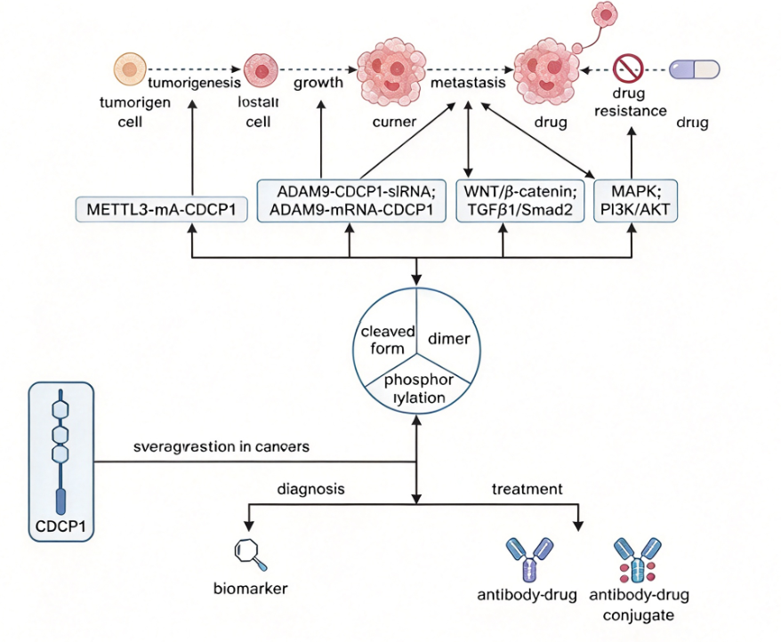
II. Proteolytic Cleavage Mechanisms and Epitope Regulation of CDCP1
Full-length 135 kDa CDCP1 is cleaved by multiple serine proteases at the adjacent Arg-368/Lys-369 sites, dividing it into a shed N-terminal fragment ATF (approximately 65 kDa) and a membrane-retained CTF (approximately 70 kDa). Which enzyme executes the cleavage depends on the system; two anchor points suffice to illustrate this complexity. The first anchor is matriptase: recombinant matriptase cleaves with high efficiency, but knocking down matriptase by approximately 90% does not alter endogenous processing——the authors' own conclusion was "efficient but non-essential." The second anchor is plasmin: in a chick chorioallantoic membrane (CAM) xenograft model of prostate cancer, it was identified as the key in vivo cleavage executor——blocking cleavage reduced metastatic focus formation on the CAM by 75% to 85%; in follow-up studies using the same strategy, cancer cell entry into the circulation through CAM blood vessels was reduced by 97% to 98%, and disseminated foci in the CAM and liver were reduced by 94% and 99%, respectively. Other executor enzymes have been identified in different systems. The accurate reading of these facts is: the cleavage site is stable, but the executor enzymes are redundant and system-dependent——any intervention pursuing a "cleavage inhibition" strategy must first answer: inhibit whom, and in which organ.
Most critically, cleavage changes the target's "face" in the eyes of antibodies——the epitope changes. Structural characterization shows that after cleavage, the N-terminal and C-terminal fragments remain tightly associated with minimal conformational change, yet the neoepitope is sufficient to be enriched by subtractive phage display: the human antibody CL03 binds only cleaved CDCP1 with a KD of 150 to 840 pM; its ADC has an EC50 of 14.1 nM in PL5 cells, extending median survival in mice from 14 days to 20–21 days, with better safety than pan-CDCP1 targeting. A contemporaneous commentary also poured cold water: the desmoplastic stroma of pancreatic cancer impedes antibody penetration, and targeting a single surface antigen may lead to immune editing and expression loss.
III. The Dual Functional Profile and Context Dependence of CDCP1
The functional literature on CDCP1 is often compressed into a single phrase——"promotes invasion and metastasis"——which is precisely the compression most in need of resistance. The first dossier is indeed substantial: most systems examined support CDCP1 promoting metastasis through the Src(Y734)→PKCδ axis——Y734F completely abolishes metastatic capacity in melanoma in vivo; among 56 primary triple-negative breast cancer specimens, the CDCP1 pY743 positivity rate in the high SFK activity group reached 100%. The drug resistance context adds another layer to this dossier: in EGFR-TKI-resistant cells, AXL/CDCP1/SRC are co-upregulated, CDCP1 silencing can partially restore sensitivity, and CDCP1 mRNA is also an independent poor prognostic factor.
The second dossier is equally solid and often overlooked. Three independent in vivo models show that CDCP1 suppresses metastasis without affecting primary tumor growth; in esophageal squamous cell carcinoma, CDCP1 mRNA is lower in tumors than in paired normal tissue, knockdown反而 increases invasion and migration, and low expressors have worse overall survival; in prostate cancer, approximately 50% of metastatic castration-resistant cases show CDCP1 loss, and CDCP1-low cells are sensitized to FAK inhibitors. Therefore, the functional direction of CDCP1 is context-dependent, and any unidirectional narrative of "CDCP1 promotes cancer, therefore target it" does not hold up. There is also an often-ignored immune dimension: in cervical cancer, CDCP1 binds T cell CD6 and suppresses cytotoxic T lymphocyte function through the CD6-JAK1 axis, with growth effects mediated by immunity——this pathway cannot be measured at all in immunodeficient models.
IV. The Therapeutic Window and Expression Profile Evidence of CDCP1
The true shape of the therapeutic window can be summarized in three statements. First statement: the tumor end is indeed broad. The six-cancer-type data come from the same study, the same antibody, and the same scoring system, making it the most uniform dataset currently available; independent cohorts each have their own criteria——lung adenocarcinoma high expression 30.0%, pancreatic cancer high expression 36.6%, triple-negative breast cancer positivity 57%, ovarian cancer high tumor score 63.72%. These numbers cannot be merged: antibody clones, scoring systems, and patient-level versus tissue-level counting all differ, and any cross-study merged positivity rate statement is imprecise. Second statement: the normal end is not blank. The 34-normal-tissue atlas provides the basis for "tumor-upregulated, normal tissue-restricted," but positivity in the hematopoietic stem/progenitor compartment and retinal pigment epithelium are clear candidate sites for off-tumor toxicity. The standard formulation should be "a tumor-upregulated, normal tissue-restricted expression profile," not "tumor-specific." Third statement: expression, prognosis, and response are three separate matters. Prognostic direction is cancer-type-dependent, and expression and response can also decouple: clear cell renal cell carcinoma shows high expression yet intrinsic resistance to MME-class ADCs. When evaluating a target with "broad expression," these three layers must be separated.
V. Conclusion
In CDCP1-related basic research and drug development, high-quality recombinant CDCP1 protein is a core tool for dissecting its antibody epitope binding mechanisms and evaluating targeted drug activity. The His tag facilitates purification via immobilized metal affinity chromatography and enables detection and capture through anti-His tag antibodies. To meet the needs of CDCP1 functional research and targeted drug screening, Uni-Ai provides CDCP1 His Tag Protein, Human. This product is suitable for applications including CDCP1 antibody epitope binding activity analysis, in vitro screening and evaluation of CDCP1-targeting antibody drugs and antibody-drug conjugates, CDCP1 proteolytic cleavage mechanism studies, and the development of tumor-targeted therapeutic strategies.
This article is reviewed and published by the technical expert team of UA
Disclaimer: This article partially utilizes artificial intelligence assistance in its creation. If any content involves copyright or intellectual property issues, please let us know and we promise to verify and remove it as soon as possible.
Purchase recombinant protein, choose 
UA protein focuses on providing various protein reagents, raw materials, and services required for drug research and development, cell therapy, gene therapy, and basic scientific research, including drug target proteins, immune checkpoint proteins, cytokines, tool enzymes, customized protein expression, and full-length transmembrane protein development. Nanjing Univ is committed to providing customers with high-quality products and professional services, and building a High-tech Biological Enterprise with International Competitiveness.
Target proteins | membrane proteins | cytokines | enzymes | viral antigens | protein customization
Nanjing UNIV Biotechnology Co., Ltd. Email:order@ua-bio.com Phone:+86-25-56221161

Product Information

Link:





©2021-2026 all rights reserved UNIV Biotechnology Co.,Ltd.
Privacy Policy
网站备案号 苏ICP备2025207586号-2 浙公网安备33010202004867号
 Business license (three certificates in one)
Tel:+86-25-56221161
E-mail:order@ua-bio.com
All products sold on this website shall not be used for clinical diagnosis or treatment of humans or animals, and shall only be used for non-medical purposes such as industrial or scientific research.


