




Cell Viability Assays: A Comprehensive Overview from Fluorescent Dyes to Chemiluminescence Technologies
Author: Senior Product Manager of UA Views:13 


This article systematically elaborates on the technical principles and methodological frameworks of cell viability assays, focusing on five major categories: fluorescent dye detection, cell metabolism detection, chemiluminescent cell activity assays, DNA synthesis detection, and cell counting assays. It analyzes the technical characteristics and applicable scenarios of these methods, as well as their differential advantages in terms of sensitivity, throughput, and operational convenience.
- Recent Advances
- Product Information
Recent Advances
Cell Viability Assays: A Comprehensive Overview from Fluorescent Dyes to Chemiluminescence
Abstract
This article systematically elaborates on the technical characteristics and application scenarios of five major categories of cell viability assays, including fluorescent dye detection, cell metabolism assays, chemiluminescent cell activity measurement, DNA synthesis detection, and cell counting assays. It analyzes the differential advantages of various detection strategies in terms of sensitivity, throughput, and operational convenience.
This article systematically elaborates on the technical characteristics and application scenarios of five major categories of cell viability assays, including fluorescent dye detection, cell metabolism assays, chemiluminescent cell activity measurement, DNA synthesis detection, and cell counting assays. It analyzes the differential advantages of various detection strategies in terms of sensitivity, throughput, and operational convenience.
1. Fundamental Concepts and Classification Framework of Cell Viability Assays
Cell viability refers to the percentage of live cells in a total cell population, where some cells may die due to various reasons. It is a critical parameter in cell biology experiments, serving as the foundation for cell culture, cytotoxicity studies, and physiological research. Based on detection principles, cell viability and proliferation assays can be categorized into five major types: fluorescent dye detection, cell metabolism assays, DNA synthesis detection, chemiluminescent cell activity measurement, and cell counting assays. Each category exhibits distinct features in sensitivity, throughput, operational convenience, and cost-effectiveness, requiring researchers to select the most suitable strategy based on experimental objectives and sample characteristics.
2. Technical Characteristics of Fluorescent Dye Detection Methods
Fluorescent dye detection methods utilize fluorescent probes to label live or dead cells, followed by quantitative analysis using flow cytometry or fluorescence microscopy. CFSE is a fluorescent dye capable of penetrating cell membranes, freely entering cells. Upon diffusion into the cell, CFSE undergoes hydrolysis by endogenous esterases, emitting green fluorescence. The fluorescent CFSE further binds to cytoskeletal proteins, forming stable intracellular fluorescent proteins. During cell division, these fluorescent proteins are evenly distributed to daughter cells, resulting in a gradual reduction in fluorescence intensity with each generation. This allows for analysis of cell proliferation via flow cytometry.
Calcein AM is one of the most ideal probes for live-cell fluorescent staining. Derived from Calcein, it incorporates an acetoxymethyl ester group to enhance hydrophobicity, facilitating membrane penetration and intracellular labeling. Upon entering the cytoplasm, esterases hydrolyze Calcein AM into Calcein, which emits green fluorescence. Dead cells, lacking or having minimal esterase activity, produce little or no Calcein and thus remain unstained. Calcein AM can be combined with propidium iodide (PI) for simultaneous staining of live and dead cells, offering advantages such as low toxicity, non-radioactivity, high throughput, and high sensitivity. Flow cytometry enables rapid and accurate detection of large cell populations, making it widely applicable for assessing proliferation status and cell viability.
3. Technical Characteristics of Cell Metabolism Assays
Cell metabolism assays indirectly reflect cell viability by measuring the metabolic activity of live cells. CCK-8 is a widely used high-sensitivity reagent kit based on WST-8 for cell proliferation and cytotoxicity assays. Similar to MTT, WST-8 is reduced by dehydrogenases in mitochondria in the presence of electron coupling reagents, generating an orange-yellow formazan product. The color intensity correlates with cell proliferation (darker color indicates higher proliferation) and cytotoxicity (lighter color indicates higher toxicity). For the same cell type, color intensity is linearly proportional to cell number. CCK-8 is extensively applied in high-throughput drug screening, cell proliferation assays, cytotoxicity tests, tumor drug sensitivity assays, live-cell counting, viability detection, and bioactivity assessment.
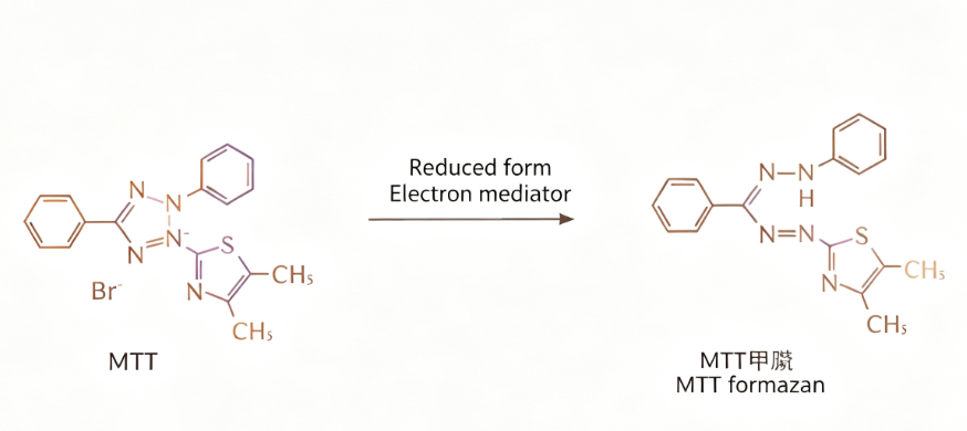
The MTT assay, also known as MTT colorimetry, relies on the reduction of exogenous MTT by succinate dehydrogenase in live cell mitochondria into insoluble purple formazan crystals, which accumulate in cells. Dead cells lack this capability. Absorbance at 490 nm is measured using a microplate reader to calculate live cell numbers. A limitation of MTT is that its reduced product is water-insoluble, requiring solubilization before detection, and it exhibits some cytotoxicity, restricting its applications. LDH release is a common cytotoxicity indicator, as LDH is normally confined to the cytoplasm but released upon membrane damage during necrosis. AlamarBlue is a next-generation cell viability indicator, where mitochondrial enzymes in live cells convert the oxidized blue form into the reduced red form, accompanied by quantifiable fluorescence changes.
4. Chemiluminescent Cell Activity Assays and DNA Synthesis Detection
ATP content measurement is the core technology of chemiluminescent cell activity assays. ATP serves as the direct energy source for cells, and the ATP luminescence method analyzes cell proliferation by detecting intracellular ATP levels. Endogenous ATP is the fundamental energy currency of living cells, rapidly hydrolyzed upon cell death. Since intracellular ATP content is positively correlated with live cell count, measuring ATP levels reflects cell viability and live cell numbers. This method offers high sensitivity, a wide linear range, and operational convenience.
DNA synthesis detection directly measures DNA synthesis in cells, considered one of the most accurate methods for assessing cell proliferation. BrdU, a thymidine analog, labels newly synthesized DNA in proliferating cells. Incorporated during DNA replication, BrdU can be detected using anti-BrdU monoclonal antibodies to evaluate proliferation capacity. EdU is a novel thymidine analog with a similar principle to BrdU, replacing thymidine in replicating DNA. By reacting with fluorescent dyes, EdU enables fluorescence-based detection of cell proliferation.
5. Cell Counting Assays
Cell counting is a fundamental method for assessing cell viability, using counting chambers or automated counters to determine cell numbers. It is simple to perform and useful for generating cell growth curves. Trypan blue staining is one of the most common methods for identifying dead cells in tissue and cell culture. This blue dye stains dead cells blue by penetrating compromised membranes, while live cells with intact membranes remain unstained. AO/PI refers to a combination of acridine orange and propidium iodide, fluorescent DNA-binding dyes emitting green and red fluorescence, respectively. AO permeates all cells, staining them green, whereas PI only enters dead cells with compromised membranes, staining nuclei red.
6. Conclusion
In practical applications of cell viability assays, high-quality detection reagents are crucial for ensuring data reliability and experimental reproducibility. Addressing this need, UA offers the UA-Glo® Luminescent Cell Viability Assay. This product is based on an ATP bioluminescence detection system and features the following core attributes: high sensitivity for quantifying ATP levels as low as femtomolar, suitable for low-cell-density viability assays; low background ensuring excellent signal-to-noise ratios; a homogeneous "add-mix-detect" workflow requiring no washing or separation steps, compatible with 96- and 384-well high-throughput formats; and stable luminescence signals supporting flexible time windows. The kit is applicable to cell proliferation assays, cytotoxicity evaluation, drug screening, and tumor drug sensitivity testing.
As a fundamental parameter in cell biology experiments, cell viability assays have evolved into a comprehensive system encompassing five major technical categories: fluorescent dyes, cell metabolism, chemiluminescence, DNA synthesis, and cell counting. Each method offers unique advantages in sensitivity, throughput, operational convenience, and cost-effectiveness, requiring researchers to select the optimal strategy based on experimental goals and sample characteristics. The UA-Glo® Luminescent Cell Viability Assay provides a reliable tool for cell viability detection, advancing research in cell biology and drug development.
This article is reviewed and published by the technical expert team of UA
Disclaimer: This article partially utilizes artificial intelligence assistance in its creation. If any content involves copyright or intellectual property issues, please let us know and we promise to verify and remove it as soon as possible.
Purchase recombinant protein, choose Nanjing UA-Bio
UA protein focuses on providing various protein reagents, raw materials, and services required for drug research and development, cell therapy, gene therapy, and basic scientific research, including drug target proteins, immune checkpoint proteins, cytokines, tool enzymes, customized protein expression, and full-length transmembrane protein development. Youai is committed to providing customers with high-quality products and professional services, and building a High-tech Biological Enterprise with International Competitiveness.
Target proteins | membrane proteins | cytokines | enzymes | viral antigens | protein customization
Nanjing UA Biotechnology Co., Ltd. Email:order@ua-bio.com Phone:+86-25-56221161

Product Information

Link:





©2021-2026 all rights reserved UA Biotechnology Co.,Ltd.
Privacy Policy 
网站备案号 浙ICP备2022019033号-2 浙公网安备33010202004867号
 Business license (three certificates in one)
 Tel:+86-25-56221161
E-mail:order@ua-bio.com
All products sold on this website shall not be used for clinical diagnosis or treatment of humans or animals, and shall only be used for non-medical purposes such as industrial or scientific research.


