




PDGF-AA: A Key Regulator of Mesenchymal Cell Proliferation and Migration
Author: Senior Product Manager of UA Views:6 


This article focuses on the molecular characteristics and biological functions of platelet-derived growth factor AA (PDGF-AA), systematically elaborating its structural features as an important ligand subtype of the PDGF family, its receptor-binding specificity, and the molecular mechanisms by which it activates signaling pathways such as PI3K/AKT and STAT3 through PDGFRα. It also analyzes its multifaceted roles in embryonic development, tissue repair, angiogenesis, and tumorigenesis.
- Recent Advances
Recent Advances
PDGF-AA: A Core Regulatory Factor in Mesenchymal Cell Proliferation and Migration
Overview
This article systematically elaborates on the molecular characteristics and biological functions of Platelet-Derived Growth Factor AA (PDGF-AA), focusing on its structural features as a key ligand subtype of the PDGF family, its receptor-binding specificity, and the molecular mechanisms by which it activates signaling pathways such as PI3K/AKT and STAT3 through PDGFRα. It also analyzes its multifaceted roles in embryonic development, tissue repair, angiogenesis, and tumorigenesis.
This article systematically elaborates on the molecular characteristics and biological functions of Platelet-Derived Growth Factor AA (PDGF-AA), focusing on its structural features as a key ligand subtype of the PDGF family, its receptor-binding specificity, and the molecular mechanisms by which it activates signaling pathways such as PI3K/AKT and STAT3 through PDGFRα. It also analyzes its multifaceted roles in embryonic development, tissue repair, angiogenesis, and tumorigenesis.
I. Molecular Composition of the PDGF Family and the Role of PDGF-AA
Platelet-Derived Growth Factor (PDGF) was the first growth factor to be identified, leading the way in the study of numerous growth factor signaling pathways and serving as a core regulator of cell growth and division. PDGF is a dimeric glycoprotein composed of two A subunits (PDGF-AA), two B subunits (PDGF-BB), or one of each (PDGF-AB). In humans and mice, the PDGF signaling network consists of five ligands: PDGF-AA, PDGF-BB, PDGF-AB, PDGF-CC, and PDGF-DD, as well as two receptors: PDGFRα and PDGFRβ. All PDGFs function as secreted, disulfide-linked homodimers, but only PDGF-A and PDGF-B can form functional heterodimers. PDGF-AA is one of the most extensively studied ligand subtypes in the PDGF family, playing an indispensable regulatory role in various physiological and pathological processes.
II. Expression Distribution and Biological Functions of PDGF-AA
PDGF-A is expressed in vascular smooth muscle cells, epithelial cells, and various tissues and organs (e.g., lung, placenta, prostate). The primary function of PDGF-AA is to promote the proliferation and migration of mesenchymal cells, participating in wound healing processes and playing critical roles in alveolar development, mesenchymal cell proliferation, gastrointestinal development, and spermatogenesis. PDGF-AA exerts mitogenic effects early in embryonic development, driving the proliferation of undifferentiated mesenchymal cells and progenitor cell populations. In mature tissues, PDGF-AA signaling is involved in tissue remodeling, cell differentiation, and inductive events related to patterning and morphogenesis. Abnormal expression of PDGF-AA is associated with malignancies such as cholangiocarcinoma and head and neck squamous cell carcinoma. Knockout studies have shown that PDGF-A-deficient mice exhibit defects in oligodendrocytes, alveolar smooth muscle cells, and testicular interstitial cells, with the mice dying during embryogenesis or within 60 days after birth, confirming the irreplaceable role of PDGF-AA in development.
III. Receptor Binding and Signal Transduction Mechanisms of PDGF-AA
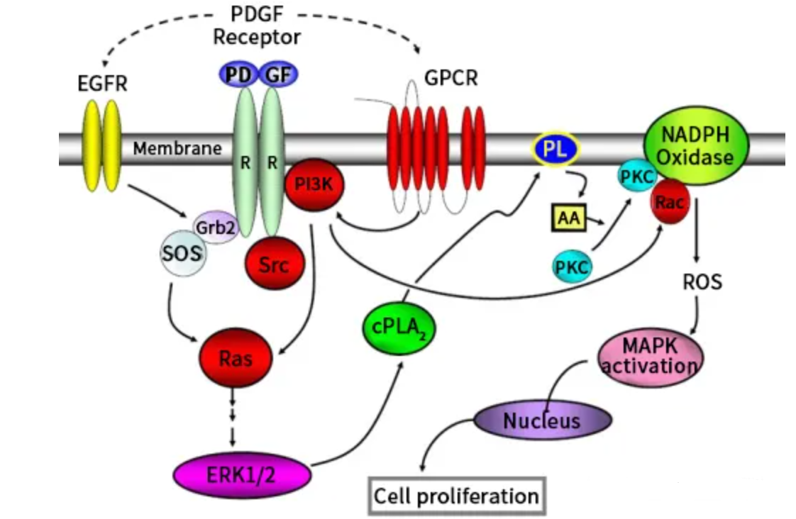
PDGF receptors are classified as receptor tyrosine kinases, with two types identified: PDGFRα and PDGFRβ. PDGFRα binds PDGF-AA, PDGF-BB, and PDGF-AB, while PDGFRβ binds PDGF-BB and PDGF-AB with high affinity. PDGF-AA is a specific ligand for PDGFRα, binding exclusively to PDGFRα and not to PDGFRβ, a selectivity that determines the specificity of PDGF-AA signaling. Upon binding to the ligand-binding pocket located within the second and third immunoglobulin domains of PDGFR, the receptor dimerizes and activates through autophosphorylation of multiple sites in the cytoplasmic domain, mediating the recruitment of auxiliary factors and the activation of downstream signaling. PDGF-AA exerts its biological effects through the PI3K/AKT pathway, the RAS/MAPK pathway, and the reactive oxygen species-mediated STAT3 pathway. In studies of mesenchymal stem cell osteogenic differentiation, PDGF-AA enhances osteogenic differentiation capacity by activating the PI3K pathway, an effect not observed with epidermal growth factor. Chemical inhibition of the PI3K pathway in PDGF-stimulated cells eliminates the differential effects between the two growth factors, confirming the central role of the PI3K pathway in the specific functions of PDGF-AA.
IV. Pathological Significance and Therapeutic Applications of PDGF-AA in Disease
Overexpression of PDGF-AA is associated with various diseases, including atherosclerosis, fibrotic disorders, and malignancies. During wound healing, PDGF-AA promotes the division and migration of fibroblasts, enabling cells to bypass the G1 checkpoint for division. Exogenous administration of PDGF stimulates the chemotaxis, proliferation, and gene expression of monocytes/macrophages and fibroblasts, significantly increasing the influx of inflammatory cells and fibroblasts and accelerating the formation of extracellular matrix and collagen. Recombinant PDGF is used medically to aid in the healing of chronic ulcers and serves as an alternative to autologous bone grafts in orthopedic surgery and periodontal disease to stimulate bone regeneration and repair.
V. Conclusion
As a ligand subtype of the PDGF family that specifically binds PDGFRα, PDGF-AA plays an indispensable central role in normal physiology and pathology through its multifaceted functions in promoting mesenchymal cell proliferation and migration, embryonic development, and tissue repair. Its involvement in tumorigenesis, fibrotic diseases, and angiogenesis makes it a critical target for therapeutic strategies in related diseases. Human recombinant PDGF-AA protein provides essential tools for in-depth analysis of the PDGF-AA/PDGFRα signaling axis and its functional networks in disease.
In PDGF-AA-related basic research and drug screening, high-quality human recombinant PDGF-AA protein is a core tool for studies on cell proliferation and migration, signaling pathway analysis, and tissue repair research. To meet these research needs, Uni offers PDGF-AA Protein, Human, suitable for applications such as mesenchymal stem cell proliferation and differentiation studies, PDGFRα signaling pathway exploration, wound healing and tissue repair experiments, and tumor microenvironment-related research.
This article is reviewed and published by the technical expert team of UA
Disclaimer: This article partially utilizes artificial intelligence assistance in its creation. If any content involves copyright or intellectual property issues, please let us know and we promise to verify and remove it as soon as possible.
Purchase recombinant protein, choose Nanjing UA-Bio
UA protein focuses on providing various protein reagents, raw materials, and services required for drug research and development, cell therapy, gene therapy, and basic scientific research, including drug target proteins, immune checkpoint proteins, cytokines, tool enzymes, customized protein expression, and full-length transmembrane protein development. Youai is committed to providing customers with high-quality products and professional services, and building a High-tech Biological Enterprise with International Competitiveness.
Target proteins | membrane proteins | cytokines | enzymes | viral antigens | protein customization
Nanjing UA Biotechnology Co., Ltd. Email:order@ua-bio.com Phone:+86-25-56221161


Link:





©2021-2026 all rights reserved UA Biotechnology Co.,Ltd.
Privacy Policy 
网站备案号 浙ICP备2022019033号-2 浙公网安备33010202004867号
 Business license (three certificates in one)
 Tel:+86-25-56221161
E-mail:order@ua-bio.com
All products sold on this website shall not be used for clinical diagnosis or treatment of humans or animals, and shall only be used for non-medical purposes such as industrial or scientific research.
