




T-cell detection for tuberculosis infection: Application and value of interferon-gamma release assays
Author: Senior Product Manager of UA Views:8 


This article focuses on the central role of gamma interferon in T-cell detection of tuberculosis infection, systematically elaborating on the immunological principles and methodological procedures of IGRA technology, and analyzing its clinical application value and limitations in latent tuberculosis screening and auxiliary diagnosis of active tuberculosis.
- Recent Advances
- Product Information
Recent Advances
T-cell Detection of Tuberculosis Infection: Application and Value of γ-Interferon Release Assay
Overview
This article focuses on the central role of γ-interferon in T-cell detection of tuberculosis infection, systematically explains the immunological principles and methodological procedures of IGRA technology, and analyzes its clinical application value and limitations in latent tuberculosis screening and auxiliary diagnosis of active tuberculosis.
This article focuses on the central role of γ-interferon in T-cell detection of tuberculosis infection, systematically explains the immunological principles and methodological procedures of IGRA technology, and analyzes its clinical application value and limitations in latent tuberculosis screening and auxiliary diagnosis of active tuberculosis.
1. Diagnostic Challenges of Tuberculosis and the Necessity of Cellular Immunity Testing.
Tuberculosis is a chronic, slow-onset infectious disease caused by Mycobacterium tuberculosis, primarily affecting the lungs but also capable of invading other organs. As a typical intracellular parasite, Mycobacterium tuberculosis primarily relies on T-cell-mediated cellular immunity rather than humoral immunity for immune responses. This characteristic directly leads to long-standing challenges in laboratory diagnosis of tuberculosis—serological methods based on antibody detection struggle to accurately reflect the protective immune status of the body, while traditional bacteriological methods such as sputum acid-fast staining and sputum culture, though highly specific, suffer from low positivity rates and long processing times, making them inadequate for rapid clinical diagnosis. The tuberculin skin test, though long-used, is severely limited in clinical value due to high false-positive rates caused by BCG vaccination and non-tuberculous mycobacterial infections. Therefore, establishing a detection method that directly reflects the immune response status of T cells to Mycobacterium tuberculosis-specific antigens has become an urgent need in the field of tuberculosis diagnosis.
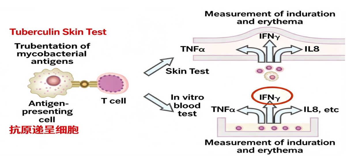
2. Immunological Principles of γ-Interferon Release Assay.
The core principle of IGRA technology is based on the biological foundation of T-cell immune memory. When the body is infected with Mycobacterium tuberculosis, specific T cells are activated and differentiate into effector memory T cells. Upon re-exposure to the same antigen, these T cells rapidly proliferate and secrete large amounts of γ-interferon, one of the primary effector molecules produced after T-cell activation. IGRA leverages this characteristic by stimulating T cells in whole blood with Mycobacterium tuberculosis-specific antigens in vitro and quantitatively measuring the concentration of IFN-γ in the culture supernatant to determine whether the subject has a specific T-cell response to Mycobacterium tuberculosis.
To achieve high specificity, the stimulating antigens used in IGRA are selected from unique regions of the pathogenic Mycobacterium tuberculosis genome, such as the RD1 and RD2 segments encoding early secretory antigen target 6 and culture filtrate protein 10. These antigens are absent in BCG strains and most non-tuberculous mycobacteria, effectively avoiding cross-reactivity interference caused by BCG vaccination and environmental mycobacterial infections.
3. Operational Procedures and Key Methodological Points of γ-Interferon Release Assay.
IGRA testing uses heparin-anticoagulated whole blood as the sample, which must be processed within 16 hours of collection. The detection system consists of three culture tubes: a background control tube (containing protein buffer to eliminate interference from inherent substances in the sample), a test culture tube (containing tuberculosis-specific stimulating antigens to induce T cells in the sample to produce and release IFN-γ), and a positive control culture tube (containing non-specific mitogens to assess the subject's basic immune function status and determine the presence of immunosuppression). After thorough mixing, the aliquoted culture tubes are incubated at 37°C for 20 to 24 hours, followed by centrifugation to separate plasma. The IFN-γ content is quantitatively measured using a double-antibody sandwich chemiluminescence method (with acridinium ester as the luminescent marker). The IFN-γ level in the positive control tube is a necessary quality control indicator to determine the validity of the test results.
4. Clinical Application Value and Interpretation of Test Results.
The primary application scenarios of IGRA technology include screening for latent tuberculosis infection, auxiliary diagnosis of active tuberculosis, and differential diagnosis of extrapulmonary tuberculosis. In immunocompromised populations (such as HIV-infected individuals), IGRA is less affected by reduced CD4+ T cells, demonstrating superior reliability compared to traditional tuberculin tests. Additionally, IGRA has a detection cycle of only 24-48 hours, high sensitivity, and is unaffected by prior BCG vaccination history, making it an important technical tool for immunological diagnosis of tuberculosis.
However, interpretation of IGRA results requires caution. A negative result does not completely rule out tuberculosis infection, as potential reasons include improper sample handling leading to cell damage, immunosuppression in the subject (such as those receiving immunosuppressive therapy or AIDS patients), or very early infection where specific T-cell memory has not yet been established. Similarly, a positive result may also be caused by rare non-tuberculous mycobacterial infections. Therefore, the diagnostic value of IGRA lies in its high negative predictive value, and positive results must still be combined with imaging, pathogen testing, and clinical manifestations for comprehensive judgment to arrive at an accurate clinical diagnosis.
5. Conclusion.
The γ-interferon release assay provides a sensitive, specific, and relatively rapid technical method for immunological diagnosis of tuberculosis infection by detecting the IFN-γ secretion response of T cells to tuberculosis-specific antigen stimulation in vitro. Clinical application of this technology requires a correct understanding of its detection principles and limitations, with results interpreted within the broader context of clinical evidence.
In T-cell detection of tuberculosis infection and γ-interferon-related basic research, high-quality IFN-γ protein is an essential tool for establishing detection methods and exploring mechanisms. To meet this research need, Uni offers IFN-γ Protein, Mouse, suitable for establishing immune detection methods for IFN-γ in mouse models, assessing cellular immune responses, and studying related signaling pathways.
This article is reviewed and published by the technical expert team of UA
Disclaimer: This article partially utilizes artificial intelligence assistance in its creation. If any content involves copyright or intellectual property issues, please let us know and we promise to verify and remove it as soon as possible.
Purchase recombinant protein, choose Nanjing UA-Bio
UA protein focuses on providing various protein reagents, raw materials, and services required for drug research and development, cell therapy, gene therapy, and basic scientific research, including drug target proteins, immune checkpoint proteins, cytokines, tool enzymes, customized protein expression, and full-length transmembrane protein development. Youai is committed to providing customers with high-quality products and professional services, and building a High-tech Biological Enterprise with International Competitiveness.
Target proteins | membrane proteins | cytokines | enzymes | viral antigens | protein customization
Nanjing UA Biotechnology Co., Ltd. Email:order@ua-bio.com Phone:+86-25-56221161

Product Information

Link:





©2021-2026 all rights reserved UA Biotechnology Co.,Ltd.
Privacy Policy 
网站备案号 浙ICP备2022019033号-2 浙公网安备33010202004867号
 Business license (three certificates in one)
 Tel:+86-25-56221161
E-mail:order@ua-bio.com
All products sold on this website shall not be used for clinical diagnosis or treatment of humans or animals, and shall only be used for non-medical purposes such as industrial or scientific research.
